Segmental Mobility
Purpose: To assess the motion at each cervical segment and the effect on the patient’s symptoms.
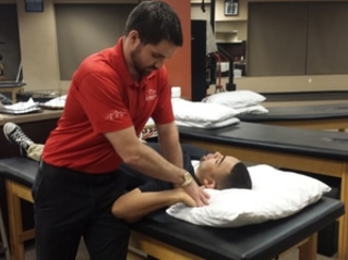
Test position: Supine.
Performing the Test: Occipitoatlantal Joint: Place both hands behind the patient’s head and rotate to the side 30 degrees. Flex and extend the head only (not the cervical joints below C1!). Repeat on the opposite side. Atlantoaxial Joint: Place both hands behind the patient’s head and flex the cervical spine maximally. Rotate the head each direction. C2-C7: Contact the articular pillar of the tested segment using the radial border of the index finger's proximal phalanx on each side. Cradle the skull using the 4th and 5th digits on each side. Flex the cervical spine maximally below the tested segment and side glide in each direction. Repeat with vertebra below tested segment in extension (the vertebrae below the test segment are flexed or extended maximally to maximally stress the capsule and joints in that direction, so that motion in other directions is limited and cannot influence the motion at the tested segment. For all of these mobility tests, compare side to side the amount of motion achieved and the pain response.
Diagnostic Accuracy: Unknown.
Importance of Test: An increase or decrease in motion in part of a segment can place additional stress on tissues that results in pain or other symptoms. This test can be used to determine how much motion is available at each segment. If a joint is restricted throughout or just unilaterally, that can aid in determining the pathology and treatment plan. By fully flexing and extending the vertebrae below, a more accurate assessment of the segmental mobility of the tested segment can be determined, because the vertebrae below are limited in motion due to full engagement of the capsule. Motion achieved during the test can be attributed mainly to the tested segment. The reason for testing in both extension and flexion is because there are potentially different restrictions in each direction that can influence movement. A thorough understanding of the spine’s osteology, arthrology, and arthrokinematics can help in understanding the results of your segmental testing. For example, in the fully flexed cervical spine, if a vertebra appears rotated to the left, that means there is a capsular restriction on that left facet. In a fully extended cervical spine, if the vertebra appears rotated to the left, that means there is a capsular restriction on the right facet.
Note: tests should only be performed by a properly trained health care practitioner.
|
|
|