- Home
- About Us
- TSPT Academy
- Online Courses
-
Resources
- Newsletter
- Business Minded Sports Physio Podcast
- Day in the Life of a Sports PT
- Residency Corner
-
Special Tests
>
-
Cervical Spine
>
- Alar Ligament Test
- Bakody's Sign
- Cervical Distraction Test
- Cervical Rotation Lateral Flexion Test
- Craniocervical Flexion Test (CCFT)
- Deep Neck Flexor Endurance Test
- Posterior-Anterior Segmental Mobility
- Segmental Mobility
- Sharp-Purser Test
- Spurling's Maneuver
- Transverse Ligament Test
- ULNT - Median
- ULNT - Radial
- ULNT - Ulnar
- Vertebral Artery Test
- Thoracic Spine >
-
Lumbar Spine/Sacroiliac Joint
>
- Active Sit-Up Test
- Alternate Gillet Test
- Crossed Straight Leg Raise Test
- Extensor Endurance Test
- FABER Test
- Fortin's Sign
- Gaenslen Test
- Gillet Test
- Gower's Sign
- Lumbar Quadrant Test
- POSH Test
- Posteroanterior Mobility
- Prone Knee Bend Test
- Prone Instability Test
- Resisted Abduction Test
- Sacral Clearing Test
- Seated Forward Flexion Test
- SIJ Compression/Distraction Test
- Slump Test
- Sphinx Test
- Spine Rotators & Multifidus Test
- Squish Test
- Standing Forward Flexion Test
- Straight Leg Raise Test
- Supine to Long Sit Test
-
Shoulder
>
- Active Compression Test
- Anterior Apprehension
- Biceps Load Test II
- Drop Arm Sign
- External Rotation Lag Sign
- Hawkins-Kennedy Impingement Sign
- Horizontal Adduction Test
- Internal Rotation Lag Sign
- Jobe Test
- Ludington's Test
- Neer Test
- Painful Arc Sign
- Pronated Load Test
- Resisted Supination External Rotation Test
- Speed's Test
- Posterior Apprehension
- Sulcus Sign
- Thoracic Outlet Tests >
- Yergason's Test
- Elbow >
- Wrist/Hand >
- Hip >
- Knee >
- Foot/Ankle >
-
Cervical Spine
>
- I want Financial Freedom
- I want Professional Growth
- I want Clinical Mastery
Alar Ligament Test
Purpose: To assess the integrity of the alar ligaments and thus upper cervical stability.
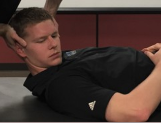
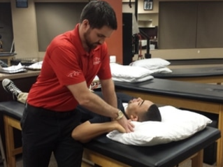
Test Position: Supine, hooklying.
Performing the Test: Place one hand on the occiput and use the other hand to palpate the spinous process of C2. Laterally flex or rotate the head to one side; you should feel the spinous process move to the opposite side. Repeat on the other side. Absence of the spinous process moving to the opposite side may indicate alar ligament injury. If you block the spinous process of C2 from moving, you may stress the ligament. You should encounter a firm end-feel in this case. Significant movement may indicate ligamentous injury.
Diagnostic Accuracy: r = .76 (“Construct validity of clinical tests for alar ligament integrity; an evaluation using magnetic resonance imaging”).
Importance of Test: Whenever a patient with neck pain as a result of trauma is being examined, you should check for alar ligament integrity. Without such testing, you could encourage a movement of the cervical spine that could damage the spinal cord. There are two alar ligaments. The distal portion of each attaches to the respective sides of the odontoid process of C2. The proximal portion attaches to the tubercle on the medial side of the respective occipital condyle. When you laterally flex or rotate the cranium to the opposite side, the atlas follows the plane of the cranium (due to ligamentous/capsular attachments). During the test, you will feel the spinous process of the axis (C2) move to the contralateral (opposite direction of the laterally flexed/rotated head), because as the proximal attachment of the alar ligament moves superiorly, the distal attachment must also move if the ligament is intact, so the spinous process is spun to the contralateral side (remember the spinal coupling that occurs int he cervical spine as well!). The alar ligament on the same side as the laterally flexed/rotated head becomes less stressed. According to Neumann, both alar ligaments are stressed during this test, but the contralateral one is stressed more. Absence of movement can indicate ligamentous instability. It should be noted that some people have a distal attachment of an anterior portion below the odontoid process or completely surpasses the odontoid process. These people may test positive for alar ligament instability. The alar ligament can have 3 directions of fiber orientation: craniocaudal, horizontal, and caudocranial. Due to this, it may be beneficial to stress the ligament in 3 planes (neutral, flexion, and extension) (“Clinical Testing for the Craniovertebral Hypermobility Syndrome”).
Note: tests should only be performed by a properly trained health care practitioner.
References:
Osmotherly PG, Rivett DA, Rowe LJ. “Construct validity of clinical tests for alar ligament integrity; an evaluation using magnetic resonance imaging.” Physical Therapy 2012 May. Epub 2012 Jan 19. Web. 08/18/2012.
Aspinall W. "Clinical Testing for the Craniovertebral Hypermobility Syndrome." Journal of Orthopedic & Sports Physical Therapy 1990. Web. 08/18/2012.
Neumann, Donald. Kinesiology of the Musculoskeletal System: Foundations for Rehabilitation. 2nd edition. St. Louis, MO: Mosby Elsevier, 2010. 336. Print.
Copyright © The Student Physical Therapist LLC 2023


