The Student​ Physical Therapist
  • Home
    • Newsletter
    • About
  • Insider Access
    • About Insider Access
  • Brian's Corner
    • Sports & Entrepreneurship Blog
    • Return to Sport Tests
    • PT Entrepreneur Course
    • Return to Sport Essentials Course
    • Become a PAID PT Consultant Course
  • Chris' Corner
  • Jim's Corner
    • Orthopedic Blog
  • Special Tests
    • Cervical Spine >
      • Alar Ligament Test
      • Bakody's Sign
      • Cervical Distraction Test
      • Cervical Rotation Lateral Flexion Test
      • Craniocervical Flexion Test (CCFT)
      • Deep Neck Flexor Endurance Test
      • Posterior-Anterior Segmental Mobility
      • Segmental Mobility
      • Sharp-Purser Test
      • Spurling's Maneuver
      • Transverse Ligament Test
      • ULNT - Median
      • ULNT - Radial
      • ULNT - Ulnar
      • Vertebral Artery Test
    • Thoracic Spine >
      • Adam's Forward Bend Test
      • Passive Neck Flexion Test
      • Thoracic Compression Test
      • Thoracic Distraction Test
      • Thoracic Foraminal Closure Test
    • Lumbar Spine/Sacroiliac Joint >
      • Active Sit-Up Test
      • Alternate Gillet Test
      • Crossed Straight Leg Raise Test
      • Extensor Endurance Test
      • FABER Test
      • Fortin's Sign
      • Gaenslen Test
      • Gillet Test
      • Gower's Sign
      • Lumbar Quadrant Test
      • POSH Test
      • Posteroanterior Mobility
      • Prone Knee Bend Test
      • Prone Instability Test
      • Resisted Abduction Test
      • Sacral Clearing Test
      • Seated Forward Flexion Test
      • SIJ Compression/Distraction Test
      • Slump Test
      • Sphinx Test
      • Spine Rotators & Multifidus Test
      • Squish Test
      • Standing Forward Flexion Test
      • Straight Leg Raise Test
      • Supine to Long Sit Test
    • Shoulder >
      • Active Compression Test
      • Anterior Apprehension
      • Biceps Load Test II
      • Drop Arm Sign
      • External Rotation Lag Sign
      • Hawkins-Kennedy Impingement Sign
      • Horizontal Adduction Test
      • Internal Rotation Lag Sign
      • Jobe Test
      • Ludington's Test
      • Neer Test
      • Painful Arc Sign
      • Pronated Load Test
      • Resisted Supination External Rotation Test
      • Speed's Test
      • Posterior Apprehension
      • Sulcus Sign
      • Thoracic Outlet Tests >
        • Adson's Test
        • Costoclavicular Brace
        • Hyperabduction Test
        • Roos (EAST)
      • Yergason's Test
    • Elbow >
      • Biceps Squeeze Test
      • Chair Sign
      • Cozen's Test
      • Elbow Extension Test
      • Medial Epicondylalgia Test
      • Mill's Test
      • Moving Valgus Stress Test
      • Push-up Sign
      • Ulnar Nerve Compression Test
      • Valgus Stress Test
      • Varus Stress Test
    • Wrist/Hand >
      • Allen's Test
      • Carpal Compression Test
      • Finkelstein Test
      • Phalen's Test
      • Reverse Phalen's Test
    • Hip >
      • Craig's Test
      • Dial Test
      • FABER Test
      • FAIR Test
      • Fitzgerald's Test
      • Hip Quadrant Test
      • Hop Test
      • Labral Anterior Impingement Test
      • Labral Posterior Impingement Test
      • Long-Axis Femoral Distraction Test
      • Noble Compression Test
      • Percussion Test
      • Sign of the Buttock
      • Trendelenburg Test
    • Knee >
      • Anterior Drawer Test
      • Dial Test (Tibial Rotation Test)
      • Joint Line Tenderness
      • Lachman Test
      • McMurray Test
      • Noble Compression Test
      • Pivot-Shift Test
      • Posterior Drawer Test
      • Posterior Sag Sign
      • Quad Active Test
      • Thessaly Test
      • Valgus Stress Test
      • Varus Stress Test
    • Foot/Ankle >
      • Anterior Drawer
      • Calf Squeeze Test
      • External Rotation Test
      • Fracture Screening Tests
      • Impingement Sign
      • Navicular Drop Test
      • Squeeze Test
      • Talar Tilt
      • Tarsal Tunnel Syndrome Test
      • Test for Interdigital Neuroma
      • Windlass Test
    • HEP >
      • Neck and Shoulder >
        • Supine Chin Tuck
        • Supine DNF with Towel Assist
        • Supine DNF
        • Standing Chin Tuck Against Wall
        • Standing Chin Tuck Against Wall with Scaption
        • Seated Cervical Retraction Repeated
        • Seated Cervical Retraction with Extension Repeated
        • Seated Cervical Retraction with Sidebend Repeated
        • Seated Cervical Retraction with Rotation Repeated
        • Standing Wall Shrugs at 90 Degrees Flex
        • Seated Thoracic Whips
        • Standing Ballistic Shoulder Extensions
        • Standing Repeated Shoulder Extension with Squat
        • Standing Repetead Shoulder Horiz. Abd. with Ext. CKC
        • Seated with Arms on Pillows Cervical AROM (Flex/Ext/Rot/SB)
        • Seated with Arms on Pillows Shrugs
        • Seated with Arms on Pillows Shrug with Scapular Retraction
        • Supine Shoulder IR with GH Centralization
        • Supine Shoulder ER with GH Centralization
        • Holding Dumbbell at 180 Degrees Flexion for Time
        • Cat Camel
        • Prone T's
        • Prone Y's
        • Quad Chin Tuck w/ Shoulder Flexion
      • Low Back >
        • Supine TA Isometric
        • Standing TA Isometric Agains Wall with Squat
        • Supine BKFO
        • Quad Rock Back
        • Standing Hip Hinge
        • Sit to Stand with Hip Hinge
        • Repeated Lumbar Sideglides
        • Repeated Standing Lumbar Extension
        • Repeated Standing Lumbar Flexion
        • Repeated Prone Press-Ups
        • Repeated Supine DKC
        • Slump Sciatic Nerve Glides
        • Birddog Progression
      • Hip and Knee >
        • Clamshells with Progressions
        • Fire Hydrants with Progressions
        • Donkey Kicks
        • Bridge Variations
        • Repeated Hip Flexion
        • Squats
        • Seated Repeated Knee Extensions
        • CKC Seated Repeated Knee Extensions
        • Heel Slides
        • CKC DF with Tibial IR
      • Foot and Ankle >
        • Calf Raises
        • Calf Raises with Soccer Ball Between Medial Malleoli
        • Towel Scrunches with Foot in PF
        • Toe Flexion Using T-Band with Foot in PF
        • PF with Toes Flexed Using T-Band
        • DF with Toes Flexed Using T-Band
        • Forefoot Adduction
        • Gastroc Stretch
        • Repeated PF
      • Examination Templates

Thoracic Spine: Anatomy Review and Clinical Implications

1/13/2014

0 Comments

 
The thoracic spine is an anatomical region that is often skimmed over in physical therapy school. We are taught basic anatomy and some assessment/treatment techniques. While this is useful, some important anatomical considerations and clinical reasoning behind when and why to use our treatment techniques is often overlooked. The thoracic spine links two of the most commonly injured body parts: cervical and lumbar spine. With the continued development of our understanding of regional interdependence, it is becoming increasingly important that we regularly use a more whole-body examination for each of our patients. The thoracic spine is a key component in this approach.
Picture
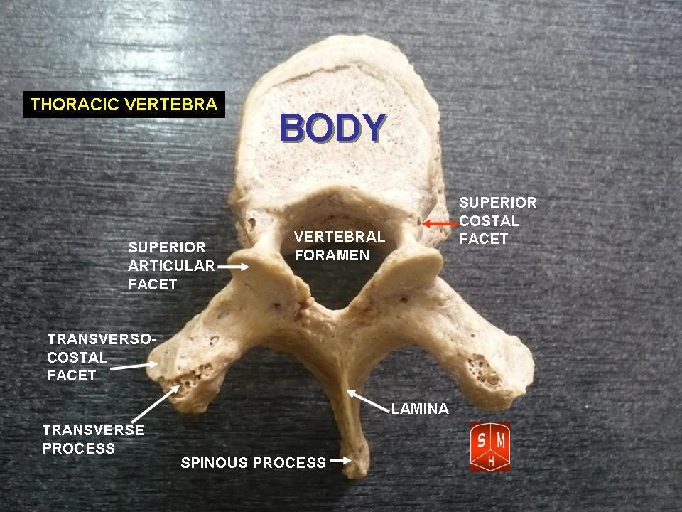
Anatomy:
One of the first things to remember is that the thoracic spine is comprised of 12 vertebrae. These vertebrae have similar characteristics of the other vertebrae: a vertebral body (disk to vertebral body height ratio is 1:5), pedicles directed posterior from the body, lamina that connect to form a spinous process, and facets (Neumann, 2010). The transverse processes are directed posterolaterally and the spinous processes fall inferiorly (depending on the region), so that they are at the level of the transverse processes below. This is an important consideration for palpation. T1-3 and T10-12 actually have their spinous processes at the same level as the transverse processes (Egan et al, 2011).  The spinous processes of T4-T6 fall half a level below the transverse processes, while T7-9 spinous processes fall a full level below the transverse processes. The pedicles actually sit directly posterior from the vertebral body, making the vertebral canal narrower here compared to other parts of the spine. T4-9 is known as the critical zone because the vertebral canal is narrowest here; it also has reduced blood supply (Egan et al, 2011). T6 is a tension point; here motion of the spinal core versus canal converge in different directions. The superior and inferior facets in the thoracic region are oriented about 60 degrees from the horizontal plane and 20 degrees from the frontal plane with the inferior facets facing anteriorly, inferiorly, and slightly medially; the superior facets face posteriorly, superiorly, and slightly lateral. Something to consider is that there is no immediate change between cervical to thoracic vertebrae and thoracic to lumbar vertebrae. The superior thoracic vertebrae bare qualities similar to the cervical spine and the inferior thoracic vertebrae resemble the lumbar spine.
Picture
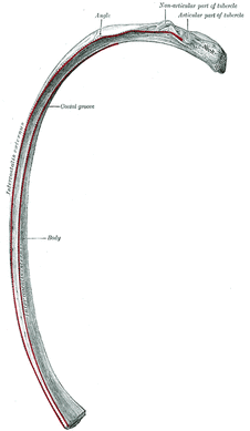
Ribs:
You cannot discuss the thoracic spine without considering the ribs as they attach in this region. There are 12 ribs (usually) in the body, just as there are 12 thoracic vertebrae. Ribs 1-7 are true ribs in that they attach directly to the sternum in the front of the body (Neumann, 2010). Ribs 8-12 are false ribs. Ribs 8-10 join in cartilage before attaching to the cartilage of the 7th rib. Ribs 11 and 12 are floating ribs and have no ventral attachments. Ribs 3-9 have demifacets on the thoracic vertebrae for attachment that span 2 vertebrae. Ribs 1, 11, and 12 have one facet on the corresponding vertebrae. The 2nd rib attaches to both T1 and T2 vertebrae. Additionally, ribs 1-10 have facet attachments at the costotransverse joints, while ribs 11-12 lack these as "floating ribs." Above T7, the rib portion of the costotransverse joints are concave, allowing for more rotation compared to the planar joints of the below T7 (Egan et al, 2011). The sympathetic chain lies on the anterior side of the rib heads next to the costovertebral joints.

Clinical Implications:
As stated previously, the fact that the thoracic spine connects the cervical spine to the lumbar spine is reason alone that this anatomical area should be considered for treatment. Think about the impact of posture. In most patients that sit for prolonged periods with a forward head posture, the anterior shift of the center of gravity places an excessive flexion moment arm to the thoracic spine, furthering the already kyphotic thoracic spine. As with any tissue, prolonged stress and creep eventually leads to pain when the tensile stiffness is no longer sufficient and the patient experiences pain. Training postural muscles is key to encouraging a more neutral posture that does not stress the tissues excessively. Consider facet restrictions: Limited joint mobility in one location often leads to hypermobility and pain in other regions, either nearby or distant. A joint dysfunction can often lead to hypertonic/painful muscle tissue near the joint as well. We can treat these restrictions with manual therapy. Two conditions less commonly discussed but seen relatively often include rib dysfunction and neural tension. Rib impairments are typically considered after a blow to the side or pain with breathing, but that is not always the case (making it harder to identify). A partially subluxed or restricted rib can be quite painful and present as thoracic paraspinal pain or even stomach pain as the tip of a floating rib presses against the anterior tissue. A potential location for pathology and treatment of neural tension is the thoracic spine. Remember that the thoracic spine contains the critical zone where the vertebral canal is narrowest. The spinal cord can easily become compressed here leading to pain/neural tension along the path of a nerve. A nerve can become irritated anywhere in the body. With the thoracic restrictions, a manipulation to the mid-thoracic spine often improves pain and neural tension by improving mobility. Think of it as a reset button as it also may have an impact on the sympathetic nervous system.

References:
Egan W, Burns S, Flynn T, and Ojha H. The Thoracic Spine and Rib Cage: Physical Therapy Patient Management Utilizing Current Evidence. Current Concepts of Orthopaedic Physical Therapy, 3rd Ed. La Crosse, WI. 2011.

Neumann, Donald. Kinesiology of the Musculoskeletal System: Foundations for Rehabilitation. 2nd edition. St. Louis, MO: Mosby Elsevier, 2010. 322-323. Print.
0 Comments

    Archives

    May 2015
    December 2014
    June 2014
    March 2014
    January 2014
    September 2013
    July 2013
    May 2013
    February 2013
    January 2013
    December 2012
    November 2012

    Categories

    All
    Elbow
    Foot/ankle
    Knee
    Research
    Shoulder
    Spine

    RSS Feed

Home

Contact Us

Copyright © The Student Physical Therapist LLC 2020
Photos used under Creative Commons from tompagenet, Iwan Gabovitch, dullhunk, hectorir, Monica's Dad, juhansonin, marc falardeau, juhansonin, brain_blogger, shock264, Menage a Moi, Wonderlane, Liz R, MateusZF Mateus Záccaro, rhodesj, hoshi7
  • Home
    • Newsletter
    • About
  • Insider Access
    • About Insider Access
  • Brian's Corner
    • Sports & Entrepreneurship Blog
    • Return to Sport Tests
    • PT Entrepreneur Course
    • Return to Sport Essentials Course
    • Become a PAID PT Consultant Course
  • Chris' Corner
  • Jim's Corner
    • Orthopedic Blog
  • Special Tests
    • Cervical Spine >
      • Alar Ligament Test
      • Bakody's Sign
      • Cervical Distraction Test
      • Cervical Rotation Lateral Flexion Test
      • Craniocervical Flexion Test (CCFT)
      • Deep Neck Flexor Endurance Test
      • Posterior-Anterior Segmental Mobility
      • Segmental Mobility
      • Sharp-Purser Test
      • Spurling's Maneuver
      • Transverse Ligament Test
      • ULNT - Median
      • ULNT - Radial
      • ULNT - Ulnar
      • Vertebral Artery Test
    • Thoracic Spine >
      • Adam's Forward Bend Test
      • Passive Neck Flexion Test
      • Thoracic Compression Test
      • Thoracic Distraction Test
      • Thoracic Foraminal Closure Test
    • Lumbar Spine/Sacroiliac Joint >
      • Active Sit-Up Test
      • Alternate Gillet Test
      • Crossed Straight Leg Raise Test
      • Extensor Endurance Test
      • FABER Test
      • Fortin's Sign
      • Gaenslen Test
      • Gillet Test
      • Gower's Sign
      • Lumbar Quadrant Test
      • POSH Test
      • Posteroanterior Mobility
      • Prone Knee Bend Test
      • Prone Instability Test
      • Resisted Abduction Test
      • Sacral Clearing Test
      • Seated Forward Flexion Test
      • SIJ Compression/Distraction Test
      • Slump Test
      • Sphinx Test
      • Spine Rotators & Multifidus Test
      • Squish Test
      • Standing Forward Flexion Test
      • Straight Leg Raise Test
      • Supine to Long Sit Test
    • Shoulder >
      • Active Compression Test
      • Anterior Apprehension
      • Biceps Load Test II
      • Drop Arm Sign
      • External Rotation Lag Sign
      • Hawkins-Kennedy Impingement Sign
      • Horizontal Adduction Test
      • Internal Rotation Lag Sign
      • Jobe Test
      • Ludington's Test
      • Neer Test
      • Painful Arc Sign
      • Pronated Load Test
      • Resisted Supination External Rotation Test
      • Speed's Test
      • Posterior Apprehension
      • Sulcus Sign
      • Thoracic Outlet Tests >
        • Adson's Test
        • Costoclavicular Brace
        • Hyperabduction Test
        • Roos (EAST)
      • Yergason's Test
    • Elbow >
      • Biceps Squeeze Test
      • Chair Sign
      • Cozen's Test
      • Elbow Extension Test
      • Medial Epicondylalgia Test
      • Mill's Test
      • Moving Valgus Stress Test
      • Push-up Sign
      • Ulnar Nerve Compression Test
      • Valgus Stress Test
      • Varus Stress Test
    • Wrist/Hand >
      • Allen's Test
      • Carpal Compression Test
      • Finkelstein Test
      • Phalen's Test
      • Reverse Phalen's Test
    • Hip >
      • Craig's Test
      • Dial Test
      • FABER Test
      • FAIR Test
      • Fitzgerald's Test
      • Hip Quadrant Test
      • Hop Test
      • Labral Anterior Impingement Test
      • Labral Posterior Impingement Test
      • Long-Axis Femoral Distraction Test
      • Noble Compression Test
      • Percussion Test
      • Sign of the Buttock
      • Trendelenburg Test
    • Knee >
      • Anterior Drawer Test
      • Dial Test (Tibial Rotation Test)
      • Joint Line Tenderness
      • Lachman Test
      • McMurray Test
      • Noble Compression Test
      • Pivot-Shift Test
      • Posterior Drawer Test
      • Posterior Sag Sign
      • Quad Active Test
      • Thessaly Test
      • Valgus Stress Test
      • Varus Stress Test
    • Foot/Ankle >
      • Anterior Drawer
      • Calf Squeeze Test
      • External Rotation Test
      • Fracture Screening Tests
      • Impingement Sign
      • Navicular Drop Test
      • Squeeze Test
      • Talar Tilt
      • Tarsal Tunnel Syndrome Test
      • Test for Interdigital Neuroma
      • Windlass Test
    • HEP >
      • Neck and Shoulder >
        • Supine Chin Tuck
        • Supine DNF with Towel Assist
        • Supine DNF
        • Standing Chin Tuck Against Wall
        • Standing Chin Tuck Against Wall with Scaption
        • Seated Cervical Retraction Repeated
        • Seated Cervical Retraction with Extension Repeated
        • Seated Cervical Retraction with Sidebend Repeated
        • Seated Cervical Retraction with Rotation Repeated
        • Standing Wall Shrugs at 90 Degrees Flex
        • Seated Thoracic Whips
        • Standing Ballistic Shoulder Extensions
        • Standing Repeated Shoulder Extension with Squat
        • Standing Repetead Shoulder Horiz. Abd. with Ext. CKC
        • Seated with Arms on Pillows Cervical AROM (Flex/Ext/Rot/SB)
        • Seated with Arms on Pillows Shrugs
        • Seated with Arms on Pillows Shrug with Scapular Retraction
        • Supine Shoulder IR with GH Centralization
        • Supine Shoulder ER with GH Centralization
        • Holding Dumbbell at 180 Degrees Flexion for Time
        • Cat Camel
        • Prone T's
        • Prone Y's
        • Quad Chin Tuck w/ Shoulder Flexion
      • Low Back >
        • Supine TA Isometric
        • Standing TA Isometric Agains Wall with Squat
        • Supine BKFO
        • Quad Rock Back
        • Standing Hip Hinge
        • Sit to Stand with Hip Hinge
        • Repeated Lumbar Sideglides
        • Repeated Standing Lumbar Extension
        • Repeated Standing Lumbar Flexion
        • Repeated Prone Press-Ups
        • Repeated Supine DKC
        • Slump Sciatic Nerve Glides
        • Birddog Progression
      • Hip and Knee >
        • Clamshells with Progressions
        • Fire Hydrants with Progressions
        • Donkey Kicks
        • Bridge Variations
        • Repeated Hip Flexion
        • Squats
        • Seated Repeated Knee Extensions
        • CKC Seated Repeated Knee Extensions
        • Heel Slides
        • CKC DF with Tibial IR
      • Foot and Ankle >
        • Calf Raises
        • Calf Raises with Soccer Ball Between Medial Malleoli
        • Towel Scrunches with Foot in PF
        • Toe Flexion Using T-Band with Foot in PF
        • PF with Toes Flexed Using T-Band
        • DF with Toes Flexed Using T-Band
        • Forefoot Adduction
        • Gastroc Stretch
        • Repeated PF
      • Examination Templates