The Student​ Physical Therapist
  • Home
  • About Us
  • Insider Access
    • About Insider Access
  • Online Courses
    • Hooper's Knee
    • Physical Therapist Entrepreneur Course
    • Physical Therapist Consultant Course
    • Orthopedic Management of the Cervical Spine
    • Return to Sport Essentials Course
  • Resources
    • Newsletter
    • Business Minded Sports Physio Podcast
    • Day in the Life of a Sports PT
    • Residency Corner
    • Special Tests >
      • Cervical Spine >
        • Alar Ligament Test
        • Bakody's Sign
        • Cervical Distraction Test
        • Cervical Rotation Lateral Flexion Test
        • Craniocervical Flexion Test (CCFT)
        • Deep Neck Flexor Endurance Test
        • Posterior-Anterior Segmental Mobility
        • Segmental Mobility
        • Sharp-Purser Test
        • Spurling's Maneuver
        • Transverse Ligament Test
        • ULNT - Median
        • ULNT - Radial
        • ULNT - Ulnar
        • Vertebral Artery Test
      • Thoracic Spine >
        • Adam's Forward Bend Test
        • Passive Neck Flexion Test
        • Thoracic Compression Test
        • Thoracic Distraction Test
        • Thoracic Foraminal Closure Test
      • Lumbar Spine/Sacroiliac Joint >
        • Active Sit-Up Test
        • Alternate Gillet Test
        • Crossed Straight Leg Raise Test
        • Extensor Endurance Test
        • FABER Test
        • Fortin's Sign
        • Gaenslen Test
        • Gillet Test
        • Gower's Sign
        • Lumbar Quadrant Test
        • POSH Test
        • Posteroanterior Mobility
        • Prone Knee Bend Test
        • Prone Instability Test
        • Resisted Abduction Test
        • Sacral Clearing Test
        • Seated Forward Flexion Test
        • SIJ Compression/Distraction Test
        • Slump Test
        • Sphinx Test
        • Spine Rotators & Multifidus Test
        • Squish Test
        • Standing Forward Flexion Test
        • Straight Leg Raise Test
        • Supine to Long Sit Test
      • Shoulder >
        • Active Compression Test
        • Anterior Apprehension
        • Biceps Load Test II
        • Drop Arm Sign
        • External Rotation Lag Sign
        • Hawkins-Kennedy Impingement Sign
        • Horizontal Adduction Test
        • Internal Rotation Lag Sign
        • Jobe Test
        • Ludington's Test
        • Neer Test
        • Painful Arc Sign
        • Pronated Load Test
        • Resisted Supination External Rotation Test
        • Speed's Test
        • Posterior Apprehension
        • Sulcus Sign
        • Thoracic Outlet Tests >
          • Adson's Test
          • Costoclavicular Brace
          • Hyperabduction Test
          • Roos (EAST)
        • Yergason's Test
      • Elbow >
        • Biceps Squeeze Test
        • Chair Sign
        • Cozen's Test
        • Elbow Extension Test
        • Medial Epicondylalgia Test
        • Mill's Test
        • Moving Valgus Stress Test
        • Push-up Sign
        • Ulnar Nerve Compression Test
        • Valgus Stress Test
        • Varus Stress Test
      • Wrist/Hand >
        • Allen's Test
        • Carpal Compression Test
        • Finkelstein Test
        • Phalen's Test
        • Reverse Phalen's Test
      • Hip >
        • Craig's Test
        • Dial Test
        • FABER Test
        • FAIR Test
        • Fitzgerald's Test
        • Hip Quadrant Test
        • Hop Test
        • Labral Anterior Impingement Test
        • Labral Posterior Impingement Test
        • Long-Axis Femoral Distraction Test
        • Noble Compression Test
        • Percussion Test
        • Sign of the Buttock
        • Trendelenburg Test
      • Knee >
        • Anterior Drawer Test
        • Dial Test (Tibial Rotation Test)
        • Joint Line Tenderness
        • Lachman Test
        • McMurray Test
        • Noble Compression Test
        • Pivot-Shift Test
        • Posterior Drawer Test
        • Posterior Sag Sign
        • Quad Active Test
        • Thessaly Test
        • Valgus Stress Test
        • Varus Stress Test
      • Foot/Ankle >
        • Anterior Drawer
        • Calf Squeeze Test
        • External Rotation Test
        • Fracture Screening Tests
        • Impingement Sign
        • Navicular Drop Test
        • Squeeze Test
        • Talar Tilt
        • Tarsal Tunnel Syndrome Test
        • Test for Interdigital Neuroma
        • Windlass Test

Exercise-related Bioenergics (aka How we use Energy!): Part 1

9/15/2013

2 Comments

 
Picture
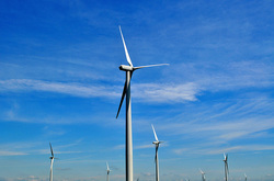
Bioenergics can be defined as "the flow of energy in a biological system, and it concerns primarily the conversion of macronutrients into biologically usable forms of energy." In other words, it is how our body utilizes energy. There are 3 main energy systems available: the phosphagen system, glycolysis, and the oxidative system. Based on the needs of a specific activity (aerobic, anaerobic, or mixed), our body selects which energy systems are required for the activity. The contribution of each system is dependent on the intensity and duration of the activity at hand. With specific exercise, each system can become more efficient and effective for a specific activity. It is for this reason, that physical therapists must understand these systems in relationship to intervention selection, dietary requirements, amount of rest, specific injury, and more to best return your client to their prior level of function (or even higher!). Part 1 is going to provide you with the background information on the physiology of each of these systems.

Picture
Back to the Basics
The energy required to perform any physical activity will either come from 1 of 2 metabolic sources: aerobic or anaerobic systems. In short, the anaerobic system is used for quick, higher intensity activities, and the aerobic system is used during longer, lower intensities activities. The energy required for us to live comes from macronutrients: carbohydrates, proteins, and fats. Each macronutrient is broken down into a measurable metabolic unit known as ATP or adenosine triphosphate. The molecule adenosine is made of adenine (a nitrogen-containing base) and ribose, a 5-carbon sugar. Triphosphate refers to 3 phosphates that are attached. In order to acquire energy from ATP,  water must be involved for hydrolysis of the molecule by adenosine triphosphatase (ATPase). So basically, energy is released when a phosphate is broken off of ATP (or ADP). The turnover rate and resynthesis of ATP depends on the intensity and duration of exercise.

Picture
The Phosphagen System
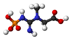
Regardless of the duration and intensity of exercise (whether sprinting 100m or running a mile), the phosphagen system is always activated at the start of exercise. Energy is derived from the previously mentioned hydrolysis of ATP and breakdown of creatine phosphate (CP). When creatine phosphate is broken down, the phosphate combines with ADP to form ATP, providing another unit of energy to be used. As you can see, ATP truly is the currency of energy. Other sources, such as CP, must be converted to ATP before they can fully be utilized. The body regularly holds about 80-100g of ATP. During exercise, levels may decrease only up to around 50%, so that energy for basic cell function can be maintained. This means as ATP is used by the body, it is being converted from other sources (such as CP or the energy stores yet to be discussed) to maintain the body's normal levels. Levels of CP in the body are usually about 4-6x higher than that of ATP. The greater the amount of Type II fibers in the body, the greater the CP storage, due to Type II fibers higher concentrations of CP. With individuals who perform brief high intensity exercise, it would make sense that they want to increase their Type II fibers, so they can have more energy for that specific activity. Unfortunately, CP is stored in small amounts and used up rapidly by the body. Phospagen stores are depleted anywhere from 1-15 (Wells 2009) seconds before the body enters glycolysis. 

Glycolysis
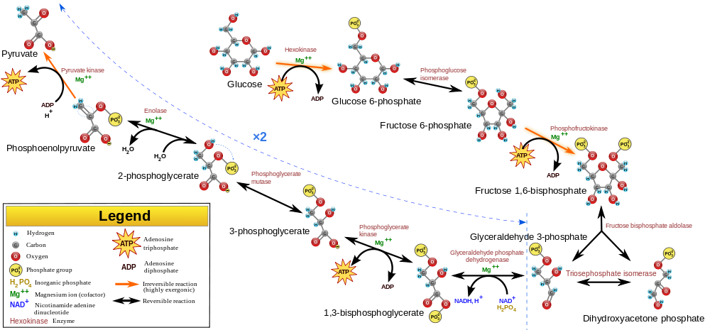
The next energy system in the line-up is glycolysis, the breakdown of carbohydrates from either glycogen in the muscle or glucose in the blood. Like CP, glycolysis' function is to create and maintain the body's normal levels of ATP. Glycolysis requires increased time to form ATP compared to CP, but it does create greater amounts. Depending on the presence of oxygen, the pyruvate either is converted to lactate or goes into the mitochondria to begin the oxidative system. If oxygen is absent and lactate forms, ATP formation speed is improved, but the amount is decreased compared to if oxygen were present. This is known as anaerobic or fast glycolysis. When oxygen is present, the pyruvate enters the Krebs cycle. This aerobic (or slow) glycolysis can last for prolonged periods, as long as the exercise intensity is low enough. If it's too high, the body relies on anaerobic means.

Lactate is often incorrectly blamed as the cause of fatigue (metabolic acidosis as a result of elevated hydrogen ion concentrations is linked to peripheral fatigue), but it does play an important role in these energy systems. It is used as an energy substrate by type I and cardiac muscle fibers. If lactate is instead transferred to the liver, it undergoes gluconeogenesis (conversion to glucose) in the Cori cycle. In fact, lactate is produced regularly, even at rest. In aerobic situations, the lactate is usually oxidized by other tissues or converted to glucose or amino acids. Clearance of lactate can be increased by light activity after exercise.

Depending on the source of glucose, glycolysis may have different ATP products. One molecule of blood glucose nets 2 ATP molecules, while muscle glycogen nets 3 ATP molecules. The difference is that blood glucose requires an additional ATP molecule to remain in its available state for energy usage. Going back to aerobic glycolysis, the pyruvate is actually transformed to Acetyl-CoA, so that it can enter the Krebs cycle. Glycolysis, like the previous system, is stimulated by elevated ADP, P, ammonia, and a decrease in pH and AMP. The opposite inhibits glycolysis. An important regulator of glycolysis is the phosphofructokinase (PFK) enzyme. With elevated ATP, PFK limits glycolysis.

As lactate begins to accumulate in the bloodstream, the sudden increase is known as the lactate threshold. It corresponds well with the ventilatory threshold as well - the point at which ventilation no longer is associated with oxygen consumption (VO2). The point at which lactate threshold occurs compared to % VO2max increases with training. A second increase in lactate levels in the bloodstream is known as onset of blood lactate accumulation (OBLA).

Picture
Oxidative System
Following aerobic glycolysis, the products enter the oxidative system. This is the energy system primarily used at rest and low intensity exercise. At rest fat is the primary source of ATP development. As exercise begins, carbohydrates become the more significant factor; however, if the exercise is prolonged with low-intensity, fats and proteins become the source of ATP development yet again. It's important to note that even when fats are the primary energy source, it is essential that a level of carbohydrates be available as well for efficient fat processing. Without adequate carbohydrate levels to facilitate the fat breakdown, it becomes much more difficult to utilize a fat for its full potential and the body is forced to breakdown protein even more.

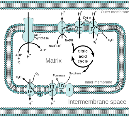
Now for a look at the cellular level of this process. Following glycolysis, the pyruvate enters the mitochondria and begins the Krebs cycle, where 2 ATP are produced for each molecule of glucose. In addition, several transport molecules (NADH and FADH2) are produced here that bring hydrogen atoms to the electron transport chain. This is where ADP becomes ATP. Oxidative phosphorylation, from the beginning of glycolysis to the end of the electron transport chain, produces 38-39 ATP. As you can see, this method produces the greatest amount of energy!
Picture
This is a very basic summary of bioenergenics, with most of the information coming from the resources listed below. Energy utilization can be broken down into much greater detail, but is beyond the purpose of this discussion. For more information about this subject, check out the sources listed below or your exercise physiology text books. Next post will discuss how we can utilize this information in designing our training programs!
References:
Baechle T & Earle R. The Essentials of Strength and Conditioning: 3rd Edition. National Strength and Conditioning      
     Association: Human Kinetics 2008. Print. 

Wells G., Selvadurai H, and Tein I. (2009). Bioenergic provision of energy for muscular activity. Paediatric Respiratory 
     Reviews. 2009; 10: 83-90. Web. 11 July 2013. 
2 Comments
Tony Lucas
7/5/2019 08:55:24 am

Hello,

I am having a hard time finding the second part to this post. Would you be able to direct me or send it to me, please?

Thank you,
Tony

Reply
Jim Heafner
8/31/2019 09:25:01 pm

Tony,

Im really sorry, but I do not think we ever followed up on this post with a Part 2. I apologize for any inconvenience, and thank you for following TSPT!

Jim

Reply


Leave a Reply.

    Archives

    May 2015
    December 2014
    June 2014
    March 2014
    January 2014
    September 2013
    July 2013
    May 2013
    February 2013
    January 2013
    December 2012
    November 2012

    Categories

    All
    Elbow
    Foot/ankle
    Knee
    Research
    Shoulder
    Spine

    RSS Feed

Home

Contact Us

Copyright © The Student Physical Therapist LLC 2022
Photos used under Creative Commons from tompagenet, Iwan Gabovitch, dullhunk, hectorir, Monica's Dad, juhansonin, marc falardeau, juhansonin, brain_blogger, shock264, Menage a Moi, Wonderlane, Liz R, MateusZF Mateus Záccaro, rhodesj, hoshi7
  • Home
  • About Us
  • Insider Access
    • About Insider Access
  • Online Courses
    • Hooper's Knee
    • Physical Therapist Entrepreneur Course
    • Physical Therapist Consultant Course
    • Orthopedic Management of the Cervical Spine
    • Return to Sport Essentials Course
  • Resources
    • Newsletter
    • Business Minded Sports Physio Podcast
    • Day in the Life of a Sports PT
    • Residency Corner
    • Special Tests >
      • Cervical Spine >
        • Alar Ligament Test
        • Bakody's Sign
        • Cervical Distraction Test
        • Cervical Rotation Lateral Flexion Test
        • Craniocervical Flexion Test (CCFT)
        • Deep Neck Flexor Endurance Test
        • Posterior-Anterior Segmental Mobility
        • Segmental Mobility
        • Sharp-Purser Test
        • Spurling's Maneuver
        • Transverse Ligament Test
        • ULNT - Median
        • ULNT - Radial
        • ULNT - Ulnar
        • Vertebral Artery Test
      • Thoracic Spine >
        • Adam's Forward Bend Test
        • Passive Neck Flexion Test
        • Thoracic Compression Test
        • Thoracic Distraction Test
        • Thoracic Foraminal Closure Test
      • Lumbar Spine/Sacroiliac Joint >
        • Active Sit-Up Test
        • Alternate Gillet Test
        • Crossed Straight Leg Raise Test
        • Extensor Endurance Test
        • FABER Test
        • Fortin's Sign
        • Gaenslen Test
        • Gillet Test
        • Gower's Sign
        • Lumbar Quadrant Test
        • POSH Test
        • Posteroanterior Mobility
        • Prone Knee Bend Test
        • Prone Instability Test
        • Resisted Abduction Test
        • Sacral Clearing Test
        • Seated Forward Flexion Test
        • SIJ Compression/Distraction Test
        • Slump Test
        • Sphinx Test
        • Spine Rotators & Multifidus Test
        • Squish Test
        • Standing Forward Flexion Test
        • Straight Leg Raise Test
        • Supine to Long Sit Test
      • Shoulder >
        • Active Compression Test
        • Anterior Apprehension
        • Biceps Load Test II
        • Drop Arm Sign
        • External Rotation Lag Sign
        • Hawkins-Kennedy Impingement Sign
        • Horizontal Adduction Test
        • Internal Rotation Lag Sign
        • Jobe Test
        • Ludington's Test
        • Neer Test
        • Painful Arc Sign
        • Pronated Load Test
        • Resisted Supination External Rotation Test
        • Speed's Test
        • Posterior Apprehension
        • Sulcus Sign
        • Thoracic Outlet Tests >
          • Adson's Test
          • Costoclavicular Brace
          • Hyperabduction Test
          • Roos (EAST)
        • Yergason's Test
      • Elbow >
        • Biceps Squeeze Test
        • Chair Sign
        • Cozen's Test
        • Elbow Extension Test
        • Medial Epicondylalgia Test
        • Mill's Test
        • Moving Valgus Stress Test
        • Push-up Sign
        • Ulnar Nerve Compression Test
        • Valgus Stress Test
        • Varus Stress Test
      • Wrist/Hand >
        • Allen's Test
        • Carpal Compression Test
        • Finkelstein Test
        • Phalen's Test
        • Reverse Phalen's Test
      • Hip >
        • Craig's Test
        • Dial Test
        • FABER Test
        • FAIR Test
        • Fitzgerald's Test
        • Hip Quadrant Test
        • Hop Test
        • Labral Anterior Impingement Test
        • Labral Posterior Impingement Test
        • Long-Axis Femoral Distraction Test
        • Noble Compression Test
        • Percussion Test
        • Sign of the Buttock
        • Trendelenburg Test
      • Knee >
        • Anterior Drawer Test
        • Dial Test (Tibial Rotation Test)
        • Joint Line Tenderness
        • Lachman Test
        • McMurray Test
        • Noble Compression Test
        • Pivot-Shift Test
        • Posterior Drawer Test
        • Posterior Sag Sign
        • Quad Active Test
        • Thessaly Test
        • Valgus Stress Test
        • Varus Stress Test
      • Foot/Ankle >
        • Anterior Drawer
        • Calf Squeeze Test
        • External Rotation Test
        • Fracture Screening Tests
        • Impingement Sign
        • Navicular Drop Test
        • Squeeze Test
        • Talar Tilt
        • Tarsal Tunnel Syndrome Test
        • Test for Interdigital Neuroma
        • Windlass Test