The Student​ Physical Therapist
  • Home
  • About Us
  • Insider Access
    • About Insider Access
  • Online Courses
    • Hooper's Knee
    • Physical Therapist Entrepreneur Course
    • Physical Therapist Consultant Course
    • Orthopedic Management of the Cervical Spine
    • Return to Sport Essentials Course
  • Resources
    • Newsletter
    • Business Minded Sports Physio Podcast
    • Day in the Life of a Sports PT
    • Residency Corner
    • Special Tests >
      • Cervical Spine >
        • Alar Ligament Test
        • Bakody's Sign
        • Cervical Distraction Test
        • Cervical Rotation Lateral Flexion Test
        • Craniocervical Flexion Test (CCFT)
        • Deep Neck Flexor Endurance Test
        • Posterior-Anterior Segmental Mobility
        • Segmental Mobility
        • Sharp-Purser Test
        • Spurling's Maneuver
        • Transverse Ligament Test
        • ULNT - Median
        • ULNT - Radial
        • ULNT - Ulnar
        • Vertebral Artery Test
      • Thoracic Spine >
        • Adam's Forward Bend Test
        • Passive Neck Flexion Test
        • Thoracic Compression Test
        • Thoracic Distraction Test
        • Thoracic Foraminal Closure Test
      • Lumbar Spine/Sacroiliac Joint >
        • Active Sit-Up Test
        • Alternate Gillet Test
        • Crossed Straight Leg Raise Test
        • Extensor Endurance Test
        • FABER Test
        • Fortin's Sign
        • Gaenslen Test
        • Gillet Test
        • Gower's Sign
        • Lumbar Quadrant Test
        • POSH Test
        • Posteroanterior Mobility
        • Prone Knee Bend Test
        • Prone Instability Test
        • Resisted Abduction Test
        • Sacral Clearing Test
        • Seated Forward Flexion Test
        • SIJ Compression/Distraction Test
        • Slump Test
        • Sphinx Test
        • Spine Rotators & Multifidus Test
        • Squish Test
        • Standing Forward Flexion Test
        • Straight Leg Raise Test
        • Supine to Long Sit Test
      • Shoulder >
        • Active Compression Test
        • Anterior Apprehension
        • Biceps Load Test II
        • Drop Arm Sign
        • External Rotation Lag Sign
        • Hawkins-Kennedy Impingement Sign
        • Horizontal Adduction Test
        • Internal Rotation Lag Sign
        • Jobe Test
        • Ludington's Test
        • Neer Test
        • Painful Arc Sign
        • Pronated Load Test
        • Resisted Supination External Rotation Test
        • Speed's Test
        • Posterior Apprehension
        • Sulcus Sign
        • Thoracic Outlet Tests >
          • Adson's Test
          • Costoclavicular Brace
          • Hyperabduction Test
          • Roos (EAST)
        • Yergason's Test
      • Elbow >
        • Biceps Squeeze Test
        • Chair Sign
        • Cozen's Test
        • Elbow Extension Test
        • Medial Epicondylalgia Test
        • Mill's Test
        • Moving Valgus Stress Test
        • Push-up Sign
        • Ulnar Nerve Compression Test
        • Valgus Stress Test
        • Varus Stress Test
      • Wrist/Hand >
        • Allen's Test
        • Carpal Compression Test
        • Finkelstein Test
        • Phalen's Test
        • Reverse Phalen's Test
      • Hip >
        • Craig's Test
        • Dial Test
        • FABER Test
        • FAIR Test
        • Fitzgerald's Test
        • Hip Quadrant Test
        • Hop Test
        • Labral Anterior Impingement Test
        • Labral Posterior Impingement Test
        • Long-Axis Femoral Distraction Test
        • Noble Compression Test
        • Percussion Test
        • Sign of the Buttock
        • Trendelenburg Test
      • Knee >
        • Anterior Drawer Test
        • Dial Test (Tibial Rotation Test)
        • Joint Line Tenderness
        • Lachman Test
        • McMurray Test
        • Noble Compression Test
        • Pivot-Shift Test
        • Posterior Drawer Test
        • Posterior Sag Sign
        • Quad Active Test
        • Thessaly Test
        • Valgus Stress Test
        • Varus Stress Test
      • Foot/Ankle >
        • Anterior Drawer
        • Calf Squeeze Test
        • External Rotation Test
        • Fracture Screening Tests
        • Impingement Sign
        • Navicular Drop Test
        • Squeeze Test
        • Talar Tilt
        • Tarsal Tunnel Syndrome Test
        • Test for Interdigital Neuroma
        • Windlass Test
Picture

Testing for Neurogenic Inhibition: Part Two (Includes a New Video from Insider Access!)

9/29/2018

0 Comments

 
Last year I wrote an article about neurogenic inhibition testing. Neurogenic Inhibition is the concept that the muscle's ability to produce resistance and contractile force is limited by the neural input, not the true muscle strength. It is indicated when either a muscle became weaker with repetitive resistance testing or if the strength improved when resistance was gradually increased. In the past, I addressed these conditions with focusing on improving mobility along the path of the nerve and with strengthening the affected muscles.
Picture
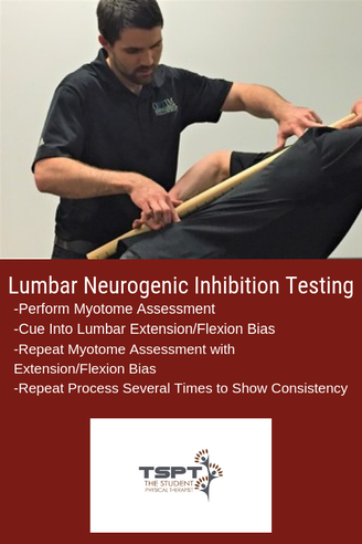
About Neurogenic Inhibition
One of my favorite aspects of my fellowship mentoring hours is that my mentor has a different treatment style and background compared to me. My training is more in line with Optim Manual Therapy's Fellowship, while my mentor went through NAIOMT's Fellowship. His coursework put a greater emphasis on testing and treating Neurogenic Inhibition. To evaluate a patient for Neurogenic Inhibition, test a patient's muscle strength with a relaxed lumbar spine, then repeat the same testing with a PPT and APT. If the strength completely normalizes with a bias of the lumbar spine, it would be positive for Neurogenic Inhibition. NAIOMT's theory is that the affected segment is "unstable" in a certain direction (decreasing the signal from the nervous system) and the lumbar spine bias provides stability that improves the neural input. An example would be supine ankle DF strength testing that was 4/5, but with the extension bias to the lumbar spine immediately becomes 5/5. The opposite may apply as well. In that same example, the 4/5 ankle DF strength may become 3+/5 with a lumbar flexion bias. It is worth testing and re-testing.

Free Preview of Insider Access!

I'm not saying that I agree with the theory, or that there is even any research to support this assessment method; however, I have noted regularly that strength changes can occur with changes in spinal positioning. The method that my mentor treats these cases is that he works on increasing strength and activation of the spine in the deficient region and then supplementing that activity with exercises that work the affected myotomal levels. For example, I evaluated a patient this past week that had 4-/5 strength of his R glutes, ankle DF and ankle eversion, all of which became 5/5 with lumbar bias into extension (complaints of drop foot for 4 years after cervical spine surgery). Some exercises we went over included ones that bias the lumbar spine into extension (APT) and work the glutes, toe extensors, and ankle eversion.

Proposed Theories 
It may be that the patients improve because of increased "stability" in the dysfunctional direction, it may instead be due to improving mobility in the dysfunctional direction, or it may be something else altogether. However, because there is so little research in the area, we don't even know how effective the method is in the long-term; however, it is worth exploring due to the immediate changes that can occur. I like to implement Neurogenic Inhibition Testing to help direct my treatment direction. I have found that this same assessment method tells me which direction a patient may respond to repeated motions. Using the same previous example, if the strength improves with lumbar extension bias, I would have the patient perform repeated lumbar extensions (or a variation of it) and recheck the strength. In most cases, the strength is improved afterwards without doing the same biasing. In fact, the patient I described came back from the evaluation with a HEP of press-ups with a R bias and his ankle DF/eversion and hip abduction strength were all 4+/5 without any lumbar spine bias. It is far too early to tell if any long-term or practical changes will occur however the testing may still play a role. It can be useful when a patient is so acute that they may not be ready for a full repeated motions assessment. In general, my treatment method is going to stay the same as discussed in the previous article: improve mobility of the nervous system along the entire path, wherever restricted, and strengthen the affected muscles. I may get there differently with this alternative testing method and I may incorporate some of the treatment theories as well.

-Dr. Chris Fox, PT, DPT, OCS

See More from The Student Physical Therapist

Picture
0 Comments

A Coper with Suspected ACL Tear: Tell Him It's Torn OR Not?

9/26/2018

0 Comments


"I was confronted with the ethical dilemma, do I tell him his ACL is likely torn or not."

​I was recently working with a 16 year old, active young man who injured his knee while playing rugby 6 days prior. During the initial evaluation, he reported quickly decelerating on the field while pivoting his body. He only had minimal pain, and his swelling was quickly improving with each day following his injury. Additionally, he had a rugby tournament overseas in 2 weeks that he needed to be in good athletic condition to play.
As I continued to the examination, I performed the Lachman's Test, which was positive, as well as a positive Anterior Drawer Test. Despite the positive finding, he denied any buckling, locking, or catching. His clinical examination was negative for meniscal pathology and other ligament insufficiency. At this point, I was confronted with the ethical dilemma: Do I tell him his ACL is likely torn or not? On one hand he was making good progress with rest and gradual return to activity. Would the diagnosis of a torn ACL create thought viruses that would hinder his progress with conservative treatment? On the other hand, if he had a torn ACL, does he potentially have other associated injuries? Segund fractures are present in 75-100% of ACL tears. Lateral meniscus tears occur in 54% of acute ACL injuries. 
Picture

Ethical Decision

As the patient, he had the right to know my clinical findings. I told him his mechanism of injury and clinical presentation are consistent with an ACL tear. Additionally, I educated him and his mother on the treatment options, copers vs. non-copers, risk of future injuries, and risks of surgery. 
The family made the decision to see an orthopedic specialist, who ordered an MRI. The MRI identified a fully torn anterior cruciate ligament and bucket handle tear of his medial meniscus. 

"The MRI identified a fully torn anterior cruciate ligament and bucket handle tear of his medial meniscus." 

The following week, he travelled with his rugby team and played in several matches. Upon his surgeon's recommendation, he would have surgery after the tournament to reconstruct his ACL and repair the meniscal tear. In this case, should surgery have been avoided? Was his ACL surgery really necessary? 

Is ACL Surgery Really Necessary? 

Picture
For many many, a torn ACL was synonymous with surgery. However recently, the optimal management of ACL injuries has been placed under question. Recent research, 'A Randomized Trial of Treatment for Acute Anterior Cruciate Ligament Tears,' identified that ACL reconstructive surgery was NOT superior to conservative management of ACL tears in young active adults. A second study by Meuffels et al, found similar outcomes between surgical and non-surgical groups at 10-year follow. The authors concluded, "we found no statistical difference between the patients treated conservatively or operatively with respect to osteoarthritis or meniscal lesions of the knee, as well as activity level, objective and subjective functional outcome."

If the outcomes are similar, why are surgeons still being performed at an alarming rate? As with most questions in physical therapy, the answer depends on a variety of factors. Clinically, I try to identify if the patient is a 'coper' or a 'non-coper.' Copers are individuals who can function at their desired level despite having a torn ACL. Non-copers are individuals who are unable to function at their prior level following an ACL injury. Copers will demonstrate good quadriceps strength, no buckling or giving way in their knee, and strong, pain free hop tests following conservative rehabilitation. Non-copers will continue to have giving way of their knee, pain that limits function, and subjective reports of decreased quality of life. Regardless, whether someone is a coper or non-coper, it is important to educate them on the pro's and con's of both surgery and rehab. 

Final Thoughts

As research continues to develop, we know at least one thing, the ACL is not vital for stability of the knee. Complications and risks will exist on either side of the equation. Patients can have success with both rehab and surgery. The job of a good physical therapist is to present the best available evidence and guide the patient in deciding which treatment option is MOST appropriate for them.
-Jim Heafner PT, DPT, OCS

Check out TSPTs NEW Knee Course!

Save $10.00 using the promo code: ACLrehab
Picture

Our Insider Access Library is Growing!

Picture
​What you get with Insider Access
 -Over 80 videos including 3 new videos every month
-Residency level education from Board Certified Orthopedic and Sports Physical Therapists
-Advanced Manual Therapy Techniques with details and descriptions from Certified Orthopedic Manual Therapists
-Return to Sport Testing and Suggested Sequencing
-Eclectic approaches to assessment and treatment
-Sport specific assessments for individual sports
References:
1. Frobell RB, Roos EM, Roos HP, Ranstam J, Lohmander LS. A randomized trial of treatment for acute anterior cruciate ligament tears. N Engl J Med2010;363:331-42.
​

2. Meuffels DE, Favejee MM, Vissers MM, Heijboer MP, Reijman M, Verhaar JA. Ten year follow-up study comparing conservative versus operative treatment of anterior cruciate ligament ruptures: a matched-pair analysis of high level athletes. Br J Sports Med2009;43:347-51.
0 Comments

ACL Return to Sport: Where are we and what are you MISSING!

9/21/2018

0 Comments

 
Picture
​Did the title of this post catch your eye? Articles with similar titles have caught my eye for years in my quest to understand proper sports rehabilitation and return to sport. Yet, despite completing a sports physical therapy residency with USC in 2014 and becoming a board certified sports physical therapist, I still find myself searching for more answers every day. I end up with more questions most of the time (which I think means I’m on the right track?).  
 
Regardless of my current quest to continue to improve my knowledge and ultimately application of knowledge in return to sport, there are a few things I have learned that are worth sharing.  Return to sport is a big buzz word and I feel confident saying that not one person has all the answers.  I’ve been lucky to be in a residency class that boasts two NFL physical therapists (Rams & Eagles) and every conversation I’ve had with them (in the past and recently) demonstrates to me that they too are constantly searching for ways to improve these processes.  Which is pretty crazy because they have great track records.  
 
My interest in ACL return to sport stems from my love for basketball and my years of special interest in treating the basketball athlete.  Unfortunately, too many basketball players suffer from ACL injuries. This sparked my interest in understanding why this happens, how we can better prepare these athletes (prevention), and what we can do to successfully return them to sport at the highest level. I say return to sport at the highest level because too often I see players return to practice level but not full game level.  
 
Currently, literature has focused on more objective criteria and milestones based progressions. However, as we know, it does take the literature time to catch up to what we see anecdotally.  Functional tests are good but do not take into consideration reactive measures.  I find myself using these tests but often adding in different movement testing with reactive components to try to mimic sports.  After all, almost all movements in sports are unplanned.  Training our athletes during their rehab or injury prevention in reactive environments can be very useful. 
 
How can we start to train “reactive” components?  I find it best not to overthink this and I often use auditory commands or visual commands. For example, when training a basketball player with shuffling in a defensive stance, I will say “Right!” or “Backwards!” or “Left!” continuously for a specified time to signal to the player to shuffle in that particular direction. Using your hands to point in specific directions is another way to do it by challenging a different sensory input. Lastly, using props such as a foam roller, tennis ball, basketball, etc to throw or drop it in a particular direction can be very effective in training reactive first steps.  It’s important to note that I often like to record these drills to look at movement both in the moment and afterwards to see what I missed with their preferred movement strategies.  
 
ACL return to sport needs to be a multifactorial approach.  As this literature article suggests, there are many ways to start preparing our athletes for their eventual return to sport.  Understanding the particular athletes sport is something that is also absolutely crucial.  Adding psychosocial components, fatigue testing, reactive testing, and sport specific movement based testing is just as important.  If you have ACL athletes and do not understand the biomechanics of their sport, take a look through the literature and check out our resources here and here.  Most importantly, continue to ask questions to yourself with each athlete you have to find continued ways to improve their outcomes. 
 
Dr. Brian Schwabe, PT, DPT, SCS, COMT, CSCS 
Board Certified Sports Physical Therapist 

​

Picture
0 Comments

Performance Hacks for Physical Therapy Evaluations

9/11/2018

4 Comments

 
Picture

What if there was ONE tool that could help you learn orthopedic evaluations as a student physical therapist (SPT)?

What if this same tool reduced errors? And was easy to use, low-tech, and cost $0?
Would you use it? What if I told you that tool was a checklist…
​When you are first learning how to perform orthopedic evaluations as an SPT, the demands on your attention can be overwhelming. Within your evaluation time frame- say you have 20-30 minutes, you must: take a subjective history, perform comprehensive testing (range of motion, strength, joint play, palpation, and special tests), as well as move the patient efficiently through multiple body positions (supine, prone, standing, sitting, etc.), and finally you must distill all of this information into a differential diagnosis. Whew! It is a lot to think about when you first start off! Any tips and tricks to improve efficiency, accuracy, and consistency of your physical therapy (PT) evaluation are truly valuable. ​
Picture
One tool that I have come to rely upon in learning how to perform a consistent and effective evaluation, in a timely manner is The Checklist. As I’ve written about before (Can A Checklist Make You A Better PT?), checklists have been used in various medical settings with positive outcomes (improving patient outcomes, reducing medical errors, and guiding treatment decisions). The checklist does not only function to reduce errors, but I believe it can be an effective learning tool for guiding appropriate practice when learning how to perform PT orthopedic evaluations.

If you want to get good at the evaluation process, it is not enough to just practice evaluations. You must take a hard look at HOW you practice evaluations. There has been a lot written about this concept in scientific journals as well as the popular literature on expertise and skill learning. (1,2,5) In order to improve a skill to the level of “mastery” or “expertise”, you must practice that skill in very specific ways. The way you practice must include focused attention as well as a means for receiving reasonably timely feedback. As students, some of this feedback can come from our professors, some can come from peers, but much of our feedback is self-delivered feedback.

​Setting up conditions for practicing evaluations is not so different from how you might approach (or teach) someone who is learning a new movement skill. The complex skill, either movement skill or orthopedic evaluation, must be broken down into its component parts and each part practiced to the extent that it is performed correctly.

Here are three key areas for optimizing your evaluation practice (and how checklists can help):

1. Break Down The Evaluation into “Sets and Reps”
     1st “Parts” Practice > 2nd “Whole” Skill Practice
  • Practice 10 “subjective” patient interviews in a row, then practice 10 “objective” patient exams in a row (if they are not all close to the “perfect” way you want them- do it again. Use the checklist to assess your accuracy/completeness).  
  • Only once your “Parts” are consistently very good should you go on to doing a full practice evaluation: practicing the “Whole”
  • If you need to break this down into smaller “Parts” to improve your consistency and quality, do it! For example: practice assessing shoulder AROM 5x in a row, until it is automatic.  
2. Increase the Quality of Your Practice Reps
     
“Practice” the way you want to “Play”
  • Well documented in the learning science literature is the idea that “practice does not make perfect. Perfect practice makes perfect”
  • Checklists can increase the “quality” of your practice reps
  • If you practice an evaluation completely “by memory” and are forgetting pieces, I hate to say it, but this is how you will most likely perform
3. Your Evaluation Should Be 75% Identical EVERY TIME
​     
The Fundamentals Must Be Automatic Before You Get Fancy
  • You need to become a robot: With the exception of special tests/add ons your flow in evaluating a certain body part should be predictable and scripted (e.g., AROM > PROM > MMT)- once this is automatic, then and only then will your higher level clinical reasoning and differential diagnosis skills improve
  • You must minimize errors of omission (leaving out key items) (Use The Checklist to make sure you have performed every piece and in the correct order)
  • You must minimize errors of commission (doing unnecessary tests), If 75% of your evaluation is the same every time- you have more cognitive resources to decide what is MOST important for that other 25%, make it count!
  • Speed/efficiency is developed second (I believe that “fast and sloppy” is never a good place to start. I tend to believe that it is best to start off with a slow but “technically” correct skill and the speed will come).
  • If you are always thinking about what comes next, you cannot effectively listen to the patient and evaluate their response (again your exam flow must be automatic). 
Picture
As clinicians, I think there is a natural desire to want to feel the state of “Flow” early on- that everything is clicking and you are utilizing a unique combination of scientific “truths” and intuitive judgments in your evaluations. But, flow and mastery take time to develop. So, I would argue that as you are starting out, the number one priority should be to make your evaluations as consistent as possible- almost to the extent of feeling “boring” or rote. This idea, I think is well expressed by famous psychologist, Mihaly Csikszentmihalyi, who coined the term “Flow” (4) and is author of the book: “Creativity: Flow and the Psychology of Discovery and Invention” (3). In this book, he writes,“You must first learn your craft and then set it aside.”

​I’m off to practice my evaluations…
 -Leda McDaniel, SPT 

Please Visit Her Website For Examples Of Her Orthopedic Evaluation Checklists

Picture
Leda is a current Doctorate of Physical Therapy (DPT) candidate at Ohio University and upon graduating in May 2019 is interested in working with orthopedic patients with chronic pain. Leda recently published a book about her experience of personal recovery from chronic pain, which you can find on Amazon:
https://www.amazon.com/dp/069212120Xref_=pe_870760_150889320
You can also find her blogging at: https://sapiensmoves.wordpress.com/


References:
  1. Brown, PC; Roediger, HL; McDaniel, MA. Make It Stick: The Science of Successful Learning. Belknap Press, 2014.
  2. Coyle, D. The Talent Code: Greatness Isn’t Born. It’s Grown. Here’s How. New York, NY. Bantam Dell; 2009.
  3. Csikszentmihalyi, M. Creativity: Flow and the Psychology of Discovery and Invention. New York, NY. HarperCollins; 2013.
  4. Csikszentmihalyi, M. Flow: The Psychology of Optimal Experience. New York, NY. HarperCollins; 1990.  
  5. Gladwell, M. Outliers: The Story of Success. New York, NY. Hachette Books; 2008. 
4 Comments
    Picture
    Dr. Brian Schwabe's NEW Book in partner with PaleoHacks!
    Picture
    Learn residency-level content on our
    Insider Access pages
    Picture

    Picture

    We value quality PT education & CEU's.  Click the MedBridge logo below for TSPT savings!


    Archives

    July 2019
    June 2019
    May 2019
    March 2019
    February 2019
    January 2019
    December 2018
    November 2018
    October 2018
    September 2018
    August 2018
    July 2018
    June 2018
    May 2018
    April 2018
    March 2018
    February 2018
    January 2018
    December 2017
    November 2017
    October 2017
    September 2017
    August 2017
    July 2017
    June 2017
    May 2017
    April 2017
    March 2017
    February 2017
    January 2017
    December 2016
    November 2016
    October 2016
    September 2016
    August 2016
    July 2016
    June 2016
    May 2016
    April 2016
    March 2016
    February 2016
    January 2016
    December 2015
    November 2015
    October 2015
    September 2015
    August 2015
    July 2015
    June 2015
    May 2015
    April 2015
    March 2015
    February 2015
    January 2015
    December 2014
    November 2014
    October 2014
    September 2014
    August 2014
    July 2014
    June 2014
    May 2014
    April 2014
    March 2014
    February 2014
    January 2014
    December 2013
    November 2013
    October 2013
    September 2013
    August 2013
    July 2013
    June 2013
    May 2013
    April 2013
    March 2013
    February 2013
    January 2013
    December 2012
    November 2012
    October 2012
    September 2012
    August 2012

    Categories

    All
    Chest
    Core Muscle
    Elbow
    Foot
    Foot And Ankle
    Hip
    Knee
    Manual Therapy
    Modalities
    Motivation
    Neck
    Neural Tension
    Other
    Research
    Research Article
    Shoulder
    Sij
    Spine
    Sports
    Therapeutic Exercise


    RSS Feed

Home

Contact Us

Copyright © The Student Physical Therapist LLC 2022
Photos used under Creative Commons from dsearls, moo.review, liverpoolhls, matturick, lwpkommunikacio, Lake Clark NPS, Tobyotter, Charly Meunier, Vincent Albanese, CNBP, Zepfanman.com, aotaro, sportEX journals, jillccarlson, Yann07, US Department of Education, osseous, TheeErin, ajy591, Mark P / PIX41, Thomas Fisher Rare Book Library, UofT, teresatrimm, donnierayjones, NNelumba, Tobyotter, Frédéric de Villamil, Jordanhill School D&T Dept, danabooo, cadillacjr2002, ingridkreuz, RDECOM, U.S. Naval Forces Central Command/U.S. Fifth Fleet, Wonderlane, hectorir, Jeffrey, Chris Hunkeler, quillons, COD Newsroom, Alan Cleaver, CCFoodTravel.com, liverpoolhls, Dr.Farouk, Jeffrey, Sten Dueland, Håkan Dahlström, PPGWings, Parker Michael Knight, MLazarevski, dno1967b, Fimb, Jim Larrison, infomatique, U.S. Naval Forces Central Command/U.S. Fifth Fleet, Idhren, iwona_kellie, AnEternalGoldenBraid, quinn.anya, jlk.1, Villainette, mlsaero, roelandpype, Defence Images, CiscoANZ, istolethetv, Our Dream Photography (Personal), BraNewbs, Meg Stewart, phalinn, akeg, hectorir, philcampbell, MilitaryHealth, akeg, Army Medicine, Shar Ka, Subconsci Productions, Eric Kilby, Anonymous9000, quinn.anya, COD Newsroom, UMN Department of Plant Pathology, familymwr, all of olive., Keith Allison, JD Hancock, Synergy by Jasmine, roger_mommaerts, John-Morgan, a.drian, Instant Vantage, Beshef, markhillary, Vegar S Hansen Photography, DEC International, kaolin fire, smith_cl9, cinnamon_girl, jillccarlson, Extra Ketchup, brain_blogger, heyjoewhereyougoinwiththatguninyourhand, tv42, COMSALUD, Nicholas_T, Ano Lobb. @healthyrx, kaibara87, mlinksva, smbuckley23, sappsnap, Menage a Moi, brain_blogger, MaloMalverde, One Way Stock, ethet., ~ggvic~, MAClarke21, Keith Allison, pictures of money, mendolus shank, HotlantaVoyeur, Mr. T in DC, abbamouse, HystericalMark, Travis Hornung, jenny downing, shock264, Peter Mooney, Futurilla, sfslim, Emily Carlin, PFX Photo, ITU Pictures, TP studio, akunamatata, Magdalena Roeseler, osseous, smith_cl9, warrenski, Sh4rp_i, mrbichel, Renaud Camus, sicamp, GreenFlames09, infomatique, mark i geo, Volker Davids, TerryJohnston, Daniela Vladimirova, CJS*64, VarsityLife, Quentin Verwaerde, Annie Mole, US Army Africa, RLHyde, openBIT e. V., Vox Efx, fabola, Gerry Dincher, mlsaero, Infomastern, N4n0, Wonderlane, TheeErin, Nanagyei, gogogadgetscott, Pulmonary Pathology, PELeCON, Elvert Barnes, @RunRockPrincess, woodleywonderworks, Asbestos Bill, Joelk75, otisarchives4, jillccarlson, sportEX journals, The Wandering Angel, perpetualplum, V31S70, colecamp, jpalinsad360, JulyYu, Andres Rueda, Whenleavesfall, melloveschallah, Pink Sherbet Photography, canonim, BrianHenry ////|//, Leonard John Matthews, mikecogh, Zepfanman.com, Kevin M. Gill, Claire L. Evans, afunkydamsel, The Official CTBTO Photostream, cyOFdevelin fame, dok1, Pam loves pie, hoshi7, opensourceway
  • Home
  • About Us
  • Insider Access
    • About Insider Access
  • Online Courses
    • Hooper's Knee
    • Physical Therapist Entrepreneur Course
    • Physical Therapist Consultant Course
    • Orthopedic Management of the Cervical Spine
    • Return to Sport Essentials Course
  • Resources
    • Newsletter
    • Business Minded Sports Physio Podcast
    • Day in the Life of a Sports PT
    • Residency Corner
    • Special Tests >
      • Cervical Spine >
        • Alar Ligament Test
        • Bakody's Sign
        • Cervical Distraction Test
        • Cervical Rotation Lateral Flexion Test
        • Craniocervical Flexion Test (CCFT)
        • Deep Neck Flexor Endurance Test
        • Posterior-Anterior Segmental Mobility
        • Segmental Mobility
        • Sharp-Purser Test
        • Spurling's Maneuver
        • Transverse Ligament Test
        • ULNT - Median
        • ULNT - Radial
        • ULNT - Ulnar
        • Vertebral Artery Test
      • Thoracic Spine >
        • Adam's Forward Bend Test
        • Passive Neck Flexion Test
        • Thoracic Compression Test
        • Thoracic Distraction Test
        • Thoracic Foraminal Closure Test
      • Lumbar Spine/Sacroiliac Joint >
        • Active Sit-Up Test
        • Alternate Gillet Test
        • Crossed Straight Leg Raise Test
        • Extensor Endurance Test
        • FABER Test
        • Fortin's Sign
        • Gaenslen Test
        • Gillet Test
        • Gower's Sign
        • Lumbar Quadrant Test
        • POSH Test
        • Posteroanterior Mobility
        • Prone Knee Bend Test
        • Prone Instability Test
        • Resisted Abduction Test
        • Sacral Clearing Test
        • Seated Forward Flexion Test
        • SIJ Compression/Distraction Test
        • Slump Test
        • Sphinx Test
        • Spine Rotators & Multifidus Test
        • Squish Test
        • Standing Forward Flexion Test
        • Straight Leg Raise Test
        • Supine to Long Sit Test
      • Shoulder >
        • Active Compression Test
        • Anterior Apprehension
        • Biceps Load Test II
        • Drop Arm Sign
        • External Rotation Lag Sign
        • Hawkins-Kennedy Impingement Sign
        • Horizontal Adduction Test
        • Internal Rotation Lag Sign
        • Jobe Test
        • Ludington's Test
        • Neer Test
        • Painful Arc Sign
        • Pronated Load Test
        • Resisted Supination External Rotation Test
        • Speed's Test
        • Posterior Apprehension
        • Sulcus Sign
        • Thoracic Outlet Tests >
          • Adson's Test
          • Costoclavicular Brace
          • Hyperabduction Test
          • Roos (EAST)
        • Yergason's Test
      • Elbow >
        • Biceps Squeeze Test
        • Chair Sign
        • Cozen's Test
        • Elbow Extension Test
        • Medial Epicondylalgia Test
        • Mill's Test
        • Moving Valgus Stress Test
        • Push-up Sign
        • Ulnar Nerve Compression Test
        • Valgus Stress Test
        • Varus Stress Test
      • Wrist/Hand >
        • Allen's Test
        • Carpal Compression Test
        • Finkelstein Test
        • Phalen's Test
        • Reverse Phalen's Test
      • Hip >
        • Craig's Test
        • Dial Test
        • FABER Test
        • FAIR Test
        • Fitzgerald's Test
        • Hip Quadrant Test
        • Hop Test
        • Labral Anterior Impingement Test
        • Labral Posterior Impingement Test
        • Long-Axis Femoral Distraction Test
        • Noble Compression Test
        • Percussion Test
        • Sign of the Buttock
        • Trendelenburg Test
      • Knee >
        • Anterior Drawer Test
        • Dial Test (Tibial Rotation Test)
        • Joint Line Tenderness
        • Lachman Test
        • McMurray Test
        • Noble Compression Test
        • Pivot-Shift Test
        • Posterior Drawer Test
        • Posterior Sag Sign
        • Quad Active Test
        • Thessaly Test
        • Valgus Stress Test
        • Varus Stress Test
      • Foot/Ankle >
        • Anterior Drawer
        • Calf Squeeze Test
        • External Rotation Test
        • Fracture Screening Tests
        • Impingement Sign
        • Navicular Drop Test
        • Squeeze Test
        • Talar Tilt
        • Tarsal Tunnel Syndrome Test
        • Test for Interdigital Neuroma
        • Windlass Test