The Student​ Physical Therapist
  • Home
  • About Us
  • Insider Access
    • About Insider Access
  • Online Courses
    • Hooper's Knee
    • Physical Therapist Entrepreneur Course
    • Physical Therapist Consultant Course
    • Orthopedic Management of the Cervical Spine
    • Return to Sport Essentials Course
  • Resources
    • Newsletter
    • Business Minded Sports Physio Podcast
    • Day in the Life of a Sports PT
    • Residency Corner
    • Special Tests >
      • Cervical Spine >
        • Alar Ligament Test
        • Bakody's Sign
        • Cervical Distraction Test
        • Cervical Rotation Lateral Flexion Test
        • Craniocervical Flexion Test (CCFT)
        • Deep Neck Flexor Endurance Test
        • Posterior-Anterior Segmental Mobility
        • Segmental Mobility
        • Sharp-Purser Test
        • Spurling's Maneuver
        • Transverse Ligament Test
        • ULNT - Median
        • ULNT - Radial
        • ULNT - Ulnar
        • Vertebral Artery Test
      • Thoracic Spine >
        • Adam's Forward Bend Test
        • Passive Neck Flexion Test
        • Thoracic Compression Test
        • Thoracic Distraction Test
        • Thoracic Foraminal Closure Test
      • Lumbar Spine/Sacroiliac Joint >
        • Active Sit-Up Test
        • Alternate Gillet Test
        • Crossed Straight Leg Raise Test
        • Extensor Endurance Test
        • FABER Test
        • Fortin's Sign
        • Gaenslen Test
        • Gillet Test
        • Gower's Sign
        • Lumbar Quadrant Test
        • POSH Test
        • Posteroanterior Mobility
        • Prone Knee Bend Test
        • Prone Instability Test
        • Resisted Abduction Test
        • Sacral Clearing Test
        • Seated Forward Flexion Test
        • SIJ Compression/Distraction Test
        • Slump Test
        • Sphinx Test
        • Spine Rotators & Multifidus Test
        • Squish Test
        • Standing Forward Flexion Test
        • Straight Leg Raise Test
        • Supine to Long Sit Test
      • Shoulder >
        • Active Compression Test
        • Anterior Apprehension
        • Biceps Load Test II
        • Drop Arm Sign
        • External Rotation Lag Sign
        • Hawkins-Kennedy Impingement Sign
        • Horizontal Adduction Test
        • Internal Rotation Lag Sign
        • Jobe Test
        • Ludington's Test
        • Neer Test
        • Painful Arc Sign
        • Pronated Load Test
        • Resisted Supination External Rotation Test
        • Speed's Test
        • Posterior Apprehension
        • Sulcus Sign
        • Thoracic Outlet Tests >
          • Adson's Test
          • Costoclavicular Brace
          • Hyperabduction Test
          • Roos (EAST)
        • Yergason's Test
      • Elbow >
        • Biceps Squeeze Test
        • Chair Sign
        • Cozen's Test
        • Elbow Extension Test
        • Medial Epicondylalgia Test
        • Mill's Test
        • Moving Valgus Stress Test
        • Push-up Sign
        • Ulnar Nerve Compression Test
        • Valgus Stress Test
        • Varus Stress Test
      • Wrist/Hand >
        • Allen's Test
        • Carpal Compression Test
        • Finkelstein Test
        • Phalen's Test
        • Reverse Phalen's Test
      • Hip >
        • Craig's Test
        • Dial Test
        • FABER Test
        • FAIR Test
        • Fitzgerald's Test
        • Hip Quadrant Test
        • Hop Test
        • Labral Anterior Impingement Test
        • Labral Posterior Impingement Test
        • Long-Axis Femoral Distraction Test
        • Noble Compression Test
        • Percussion Test
        • Sign of the Buttock
        • Trendelenburg Test
      • Knee >
        • Anterior Drawer Test
        • Dial Test (Tibial Rotation Test)
        • Joint Line Tenderness
        • Lachman Test
        • McMurray Test
        • Noble Compression Test
        • Pivot-Shift Test
        • Posterior Drawer Test
        • Posterior Sag Sign
        • Quad Active Test
        • Thessaly Test
        • Valgus Stress Test
        • Varus Stress Test
      • Foot/Ankle >
        • Anterior Drawer
        • Calf Squeeze Test
        • External Rotation Test
        • Fracture Screening Tests
        • Impingement Sign
        • Navicular Drop Test
        • Squeeze Test
        • Talar Tilt
        • Tarsal Tunnel Syndrome Test
        • Test for Interdigital Neuroma
        • Windlass Test
Picture

Exploring the Unknown

11/26/2016

0 Comments

 
Picture
At least once a year I make an attempt to argue for the benefit of reaching beyond the current clinical standards. There are many who live and die by a single pillar of evidence-based practice: research. While it is an essential component to improve our practice patterns, it has limitations. Some of the crazier techniques out there are currently impossible to standardize and accurately assess. Should this alone make them useless? The other pillars include patient beliefs and clinical experience. With the power the mind plays in pain and dysfunction, it is essential we do whatever it takes to help our patient, even if the higher level research doesn't support it.

Am I saying we should abandon what decades of research have taught us? Absolutely not. This evidence should absolutely guide our decision-making, just not rule it. For example, should a patient come in with patellafemoral pain syndrome, the evidence says we should incorporate quadriceps and gluteus strengthening. However, if tibial IR mobility is limited, we may possibly significantly improve the patient's function and pain through simply addressing that. There is no research to support this concept but has been seen clinically by many clinicians who implement repeated motions.

Even repeated motions has some sort of foundation of research. There are other techniques and schools of practice out there that are laughed at and have shown significant clinical success, such as visceral treatment, craniosacral, dry needling and more. I'm not sure these techniques can be categorized the same way much of the EBP followers are used to, but success can be shown with implementation of asterisk signs (even if we don't know the mechanism). Without some individuals attempting to go outside the current boundaries of evidence-based practice, we would fail to learn not only what techniques or treatment styles work, but also what doesn't work.

-Dr. Chris Fox, PT, DPT, OCS


If you are looking to improve upon your clinical skills, orthopaedic knowledge and clinical decision making, consider joining OPTIM's COMT program. With OPTIM, you can expect a residency-like learning experience without breaking the bank, all while learning from highly skilled physical therapists. Check out optimfellowship.com for more information!
Picture

Picture
0 Comments

The Importance of a Standardized Mobility Exam

11/19/2016

3 Comments

 
Picture
Regional interdependence is a concept regularly being taught currently, that describes how a limitation in one region can affect the movement and pain in another region. For example, limited thoracic rotation may limit overall shoulder elevation mobility and result in excessive wear/pain. With a standardized mobility examination (like the Selective Functional Movement Assessment - SFMA), we are better able to pick up some of these deficiencies.

While a system like the SFMA is built more on musculoskeletal limitations, it can be useful for other systems as well. Some of you may have seen my post a couple weeks ago on my experience with visceral therapy. It is said that there is a visceral component in 80% of musculoskeletal injuries. Some of my thoracic extension mobility restrictions were secondary to visceral issues. The concept makes sense generally speaking if you think about how any tissue can theoretically resist motion. With referral patterns for pain as well, they can present with a sort of pattern. That doesn't mean visceral restrictions cannot improve without visceral treatment. However, it does mean we need to be extremely thorough with our examination. If cervical mobility is limited due to a restriction in the liver, there will unlikely be much improvement if only the neck and upper quarter observed. A system like the SFMA forces you to look at the entire body and possibly have some success through treatment. In being forced to be thorough, you will be much less likely to miss significant findings, that at first glance, appear minimal.

Be sure to check out my post on my experience with visceral therapy from a couple weeks ago. I'm excited to be signed up or the level 1 course in April! Check out www.barralinstitute.com for more details on their methods of treatment/evaluation.

-Dr. Chris Fox, PT, DPT, OCS


If you are looking to improve upon your clinical skills, orthopaedic knowledge and clinical decision making, consider joining OPTIM's COMT program. With OPTIM, you can expect a residency-like learning experience without breaking the bank, all while learning from highly skilled physical therapists. Check out optimfellowship.com for more information!
Picture
Picture
3 Comments

Gait Assessment Clinical Short Cuts

11/16/2016

1 Comment

 
Picture
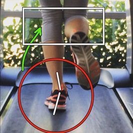
Whether a physical therapist is performing a cervical evaluation or ankle evaluation, analyzing a patient's gait pattern should be a standard aspect of every examination. The information gathered during this functional assessment will allow the clinician to quickly make educated decisions regarding the patient's overall presentation. For example, if the patient has a compensated Trendelenburg during the stance phase of gait, one can assume the strength or motor control of the Gluteus Medius is insufficient. One's ability to recognize these patterns will ultimately differentiate a novice clinician from an expert.   

Gait Analysis Considerations
Each statement below can be due to a number of reasons. I have chosen to include some of the more common clinical short cuts I use. Feel free to add others in the comment box.

1) Weight bearing equally through both legs
2) Equal and adequate hip extension bilaterally

If the patient does not demonstrate adequate hip extension while ambulating, it is important to assess the strength of the gluteals and/or length of the hip flexors. Clinically, strong/dominant hip flexors will limit a patient's full hip extension.  
3) Trunk control during single limb loading
While watching a patient walk, does the torso remain relatively neutral? A common fault often seen in patient's with low back pain is excessive pelvic motion or rotation through the spine. This impairment may indicate poor motor control of the core muscles or poor lumbopelvic rhythm. If this is noted, further motor control testing should be performed.  
4) Lacking ankle and knee motion
If a patient lacks ankle dorsiflexion, they may compensate by out-toeing their foot or going into an early heel rise. In either situation, the gastroc muscle is not being utilized throughout the full range of motion. The compensation at the ankle can create achilles issues, knee pain due to the added stress on the quadriceps muscle, or low back pain.  
5) Trendelenburg or compensated Trendelenburg
The ability to remain upright through the hips will demonstrate the strength or motor control of the outer hip muscles. When either a Trendelenburg or compensated Trendelenburg is observed, it is important to assess gluteus medius strength. Additionally, this sign can be an indication to assess lumbar mobility, specifically lumbar side bending. The poor gluteal performance will often cause the low back to load asymmetrically with each step. 
6) Knee valgus during single limb stance
Observing knee valgus during the gait assessment is another indication for assessing gluteal strength. Knee valgus is typically accompanied by femoral internal rotation and adduction, both signs of inadequate gluteal muscle or hip external rotation activation. Additionally, knee valgus is common in people who over pronate. 
7) Excessive pronation or supination
Since I mentioned pronation above,  it is important to monitor for excessive supination as well. When supination is noted throughout the gait pattern, it may indicate joint mobility restrictions in great toe extension, subtalar eversion and/or restrictions in hip internal and external range of motion.  
Whenever analyzing one's gait pattern, be sure to be systematic. Either perform the assessment from top-down or vice versa. Look for common substitutions first, then verify these impairments during your joint-by-joint examination! Identifying these common movement substitutions will make you a more efficient clinician. Repeated clinical efficiency will create excellence! 

Jim Heafner PT, DPT, OCS
Picture
Want to learn more about efficiency? Check out Jim's ebook, "The Guide to Efficient Physical Therapy Examination." This book breaks down concepts similar to the gait analysis post above to help clinicians with pattern recognition.  Speed up your evaluation and give yourself more time to treat! 
Use code: TSPT50 to save >$20 on the book. 

Picture
OPTIM Manual Therapy is now enrolling the Dallas, TX cohort for the COMT program. OPTIM's unique curriculum provides participants a well-rounded, “Monday morning applicable” experience. OPTIM was started with one singular goal in mind. We want to improve the practice of physical therapy. We promote excellence in psychomotor skills, exercise prescription, differential diagnosis, and pain science. Please contact OPTIM if you are interested! 

1 Comment

Mini-Case: Lateral Foot Pain

11/14/2016

0 Comments

 
Picture
Last week, I was treating another PT's patient with the diagnosis of patellafemoral pain syndrome (PFPS). On this day she was complaining of L lateral foot pain after having to wear heels a couple times during the week. With the location of pain, we should immediately develop multiple hypotheses. The patient's asterisk sign was lateral foot pain/tightness with lumbar flexion while in a half lateral split position.

Initial hypotheses based on pain position could suggest cuboid dysfunction, S1 radiculopathy, Intermediate Dorsal Cutaneous neural tension, peroneal strain, lateral ankle sprain, or more. I immediately found no local pain with palpation or muscle testing (making these diagnoses less likely), so my mind went to the spine. I did have some mild pain recreation with common peroneal neural tensioning, but it wasn't a significant amount. While the patient did have some lumbar mobility restrictions, mobilizing the lumbar spine and performing repeated motions had limited improvement in the asterisk sign. Even though I typically like to start at the spine with treatment, some cases are more peripherally based.  At this point I addressed the proximal tib-fib joint restriction due to the proximity of the common peroneal nerve, which has a distal branch known as the Intermediate Dorsal Cutaneous nerve. Still no significant change in the asterisk sign occurred.

Fortunately, earlier this year I took a course on dry needling. One of the things I found most beneficial in the course was learning about trigger point referral patterns. A commonly involved muscle, and the culprit in this case, is the gluteus minimus. While the muscle frequently presents with local pain and a "sciatica-like" distribution, it can also present with lateral foot pain (based on the trigger point charts in our dry needling workbooks). After needling this muscle, the patient's asterisk sign significantly improved and the patient returned this week with no more pain in that foot. The lesson from a case like this is that we should not become overly focused on one body region. While many injuries will respond to treatment through different methods, there are certain injuries that require specific treatment.

-Dr. Chris Fox, PT, DPT, OCS

If you are looking to improve upon your clinical skills, orthopaedic knowledge and clinical decision making, consider joining OPTIM's COMT program. With OPTIM, you can expect a residency-like learning experience without breaking the bank, all while learning from highly skilled physical therapists. Check out optimfellowship.com for more information!
Picture

Picture
0 Comments

Stopping Bad Habits as a Clinician

11/7/2016

0 Comments

 
We are frequently taught that we should be able to get about 80% of the information we need to figure what is wrong with a patient, simply through our subjective evaluation. What is frequently missed, however, is what we are telling patients (verbally and non-verbally) and the impact it has. Mike Reinold recently wrote an article on 6 things we shouldn't do as physical therapists. It is definitely worth a read. There are things we do as therapists that we may not realize the impact. With the development of pain science research, we are learning that the things we say and how we act impact patients significantly. This includes the diagnostic process. It is best to avoid using pathoanatomical wording, as it creates a fearful and potentially hopeless attitude in the patient. Focus more on what we can do as physical therapists, i.e. “get your hip moving,” “strengthen these muscles,” and “teach you how to move.” Find out what the patient wants out of therapy and work to align your goals with theirs. It is essential that we establish a two-way relationship with our patients so that trust can be the foundation for improvement.

-Dr. Chris Fox, PT, DPT, OCS

If you are looking to improve upon your clinical skills, orthopaedic knowledge and clinical decision making, consider joining OPTIM's COMT program. With OPTIM, you can expect a residency-like learning experience without breaking the bank, all while learning from highly skilled physical therapists. Check out optimfellowship.com for more information!
Picture

Picture
0 Comments

Insider Access Preview: Screening for Visceral Involvement

11/1/2016

0 Comments

 
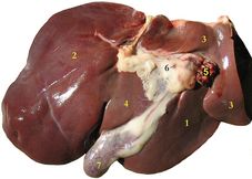
For October's Insider Access Post, I wanted to make the video available to all due to the little known importance of the topic. As many of you saw in my post a couple weeks ago, I had an incredible response to visceral treatment for an odd symptom of throat tightness. According to the therapists that treated me, there can be visceral involvement in orthopaedic injuries up to 80% of the time. Many of the patients that don't respond to our treatment, may benefit from seeing a visceral therapist. Below is a video on how to help screen for these patients. Visceral pain referral patterns are helpful for this, but insufficient.

-Chris Fox, PT, DPT, OCS
0 Comments
    Picture
    Dr. Brian Schwabe's NEW Book in partner with PaleoHacks!
    Picture
    Learn residency-level content on our
    Insider Access pages
    Picture

    Picture

    We value quality PT education & CEU's.  Click the MedBridge logo below for TSPT savings!


    Archives

    July 2019
    June 2019
    May 2019
    March 2019
    February 2019
    January 2019
    December 2018
    November 2018
    October 2018
    September 2018
    August 2018
    July 2018
    June 2018
    May 2018
    April 2018
    March 2018
    February 2018
    January 2018
    December 2017
    November 2017
    October 2017
    September 2017
    August 2017
    July 2017
    June 2017
    May 2017
    April 2017
    March 2017
    February 2017
    January 2017
    December 2016
    November 2016
    October 2016
    September 2016
    August 2016
    July 2016
    June 2016
    May 2016
    April 2016
    March 2016
    February 2016
    January 2016
    December 2015
    November 2015
    October 2015
    September 2015
    August 2015
    July 2015
    June 2015
    May 2015
    April 2015
    March 2015
    February 2015
    January 2015
    December 2014
    November 2014
    October 2014
    September 2014
    August 2014
    July 2014
    June 2014
    May 2014
    April 2014
    March 2014
    February 2014
    January 2014
    December 2013
    November 2013
    October 2013
    September 2013
    August 2013
    July 2013
    June 2013
    May 2013
    April 2013
    March 2013
    February 2013
    January 2013
    December 2012
    November 2012
    October 2012
    September 2012
    August 2012

    Categories

    All
    Chest
    Core Muscle
    Elbow
    Foot
    Foot And Ankle
    Hip
    Knee
    Manual Therapy
    Modalities
    Motivation
    Neck
    Neural Tension
    Other
    Research
    Research Article
    Shoulder
    Sij
    Spine
    Sports
    Therapeutic Exercise


    RSS Feed

Home

Contact Us

Copyright © The Student Physical Therapist LLC 2022
Photos used under Creative Commons from dsearls, moo.review, liverpoolhls, matturick, lwpkommunikacio, Lake Clark NPS, Tobyotter, Charly Meunier, Vincent Albanese, CNBP, Zepfanman.com, aotaro, sportEX journals, jillccarlson, Yann07, US Department of Education, osseous, TheeErin, ajy591, Mark P / PIX41, Thomas Fisher Rare Book Library, UofT, teresatrimm, donnierayjones, NNelumba, Tobyotter, Frédéric de Villamil, Jordanhill School D&T Dept, danabooo, cadillacjr2002, ingridkreuz, RDECOM, U.S. Naval Forces Central Command/U.S. Fifth Fleet, Wonderlane, hectorir, Jeffrey, Chris Hunkeler, quillons, COD Newsroom, Alan Cleaver, CCFoodTravel.com, liverpoolhls, Dr.Farouk, Jeffrey, Sten Dueland, Håkan Dahlström, PPGWings, Parker Michael Knight, MLazarevski, dno1967b, Fimb, Jim Larrison, infomatique, U.S. Naval Forces Central Command/U.S. Fifth Fleet, Idhren, iwona_kellie, AnEternalGoldenBraid, quinn.anya, jlk.1, Villainette, mlsaero, roelandpype, Defence Images, CiscoANZ, istolethetv, Our Dream Photography (Personal), BraNewbs, Meg Stewart, phalinn, akeg, hectorir, philcampbell, MilitaryHealth, akeg, Army Medicine, Shar Ka, Subconsci Productions, Eric Kilby, Anonymous9000, quinn.anya, COD Newsroom, UMN Department of Plant Pathology, familymwr, all of olive., Keith Allison, JD Hancock, Synergy by Jasmine, roger_mommaerts, John-Morgan, a.drian, Instant Vantage, Beshef, markhillary, Vegar S Hansen Photography, DEC International, kaolin fire, smith_cl9, cinnamon_girl, jillccarlson, Extra Ketchup, brain_blogger, heyjoewhereyougoinwiththatguninyourhand, tv42, COMSALUD, Nicholas_T, Ano Lobb. @healthyrx, kaibara87, mlinksva, smbuckley23, sappsnap, Menage a Moi, brain_blogger, MaloMalverde, One Way Stock, ethet., ~ggvic~, MAClarke21, Keith Allison, pictures of money, mendolus shank, HotlantaVoyeur, Mr. T in DC, abbamouse, HystericalMark, Travis Hornung, jenny downing, shock264, Peter Mooney, Futurilla, sfslim, Emily Carlin, PFX Photo, ITU Pictures, TP studio, akunamatata, Magdalena Roeseler, osseous, smith_cl9, warrenski, Sh4rp_i, mrbichel, Renaud Camus, sicamp, GreenFlames09, infomatique, mark i geo, Volker Davids, TerryJohnston, Daniela Vladimirova, CJS*64, VarsityLife, Quentin Verwaerde, Annie Mole, US Army Africa, RLHyde, openBIT e. V., Vox Efx, fabola, Gerry Dincher, mlsaero, Infomastern, N4n0, Wonderlane, TheeErin, Nanagyei, gogogadgetscott, Pulmonary Pathology, PELeCON, Elvert Barnes, @RunRockPrincess, woodleywonderworks, Asbestos Bill, Joelk75, otisarchives4, jillccarlson, sportEX journals, The Wandering Angel, perpetualplum, V31S70, colecamp, jpalinsad360, JulyYu, Andres Rueda, Whenleavesfall, melloveschallah, Pink Sherbet Photography, canonim, BrianHenry ////|//, Leonard John Matthews, mikecogh, Zepfanman.com, Kevin M. Gill, Claire L. Evans, afunkydamsel, The Official CTBTO Photostream, cyOFdevelin fame, dok1, Pam loves pie, hoshi7, opensourceway
  • Home
  • About Us
  • Insider Access
    • About Insider Access
  • Online Courses
    • Hooper's Knee
    • Physical Therapist Entrepreneur Course
    • Physical Therapist Consultant Course
    • Orthopedic Management of the Cervical Spine
    • Return to Sport Essentials Course
  • Resources
    • Newsletter
    • Business Minded Sports Physio Podcast
    • Day in the Life of a Sports PT
    • Residency Corner
    • Special Tests >
      • Cervical Spine >
        • Alar Ligament Test
        • Bakody's Sign
        • Cervical Distraction Test
        • Cervical Rotation Lateral Flexion Test
        • Craniocervical Flexion Test (CCFT)
        • Deep Neck Flexor Endurance Test
        • Posterior-Anterior Segmental Mobility
        • Segmental Mobility
        • Sharp-Purser Test
        • Spurling's Maneuver
        • Transverse Ligament Test
        • ULNT - Median
        • ULNT - Radial
        • ULNT - Ulnar
        • Vertebral Artery Test
      • Thoracic Spine >
        • Adam's Forward Bend Test
        • Passive Neck Flexion Test
        • Thoracic Compression Test
        • Thoracic Distraction Test
        • Thoracic Foraminal Closure Test
      • Lumbar Spine/Sacroiliac Joint >
        • Active Sit-Up Test
        • Alternate Gillet Test
        • Crossed Straight Leg Raise Test
        • Extensor Endurance Test
        • FABER Test
        • Fortin's Sign
        • Gaenslen Test
        • Gillet Test
        • Gower's Sign
        • Lumbar Quadrant Test
        • POSH Test
        • Posteroanterior Mobility
        • Prone Knee Bend Test
        • Prone Instability Test
        • Resisted Abduction Test
        • Sacral Clearing Test
        • Seated Forward Flexion Test
        • SIJ Compression/Distraction Test
        • Slump Test
        • Sphinx Test
        • Spine Rotators & Multifidus Test
        • Squish Test
        • Standing Forward Flexion Test
        • Straight Leg Raise Test
        • Supine to Long Sit Test
      • Shoulder >
        • Active Compression Test
        • Anterior Apprehension
        • Biceps Load Test II
        • Drop Arm Sign
        • External Rotation Lag Sign
        • Hawkins-Kennedy Impingement Sign
        • Horizontal Adduction Test
        • Internal Rotation Lag Sign
        • Jobe Test
        • Ludington's Test
        • Neer Test
        • Painful Arc Sign
        • Pronated Load Test
        • Resisted Supination External Rotation Test
        • Speed's Test
        • Posterior Apprehension
        • Sulcus Sign
        • Thoracic Outlet Tests >
          • Adson's Test
          • Costoclavicular Brace
          • Hyperabduction Test
          • Roos (EAST)
        • Yergason's Test
      • Elbow >
        • Biceps Squeeze Test
        • Chair Sign
        • Cozen's Test
        • Elbow Extension Test
        • Medial Epicondylalgia Test
        • Mill's Test
        • Moving Valgus Stress Test
        • Push-up Sign
        • Ulnar Nerve Compression Test
        • Valgus Stress Test
        • Varus Stress Test
      • Wrist/Hand >
        • Allen's Test
        • Carpal Compression Test
        • Finkelstein Test
        • Phalen's Test
        • Reverse Phalen's Test
      • Hip >
        • Craig's Test
        • Dial Test
        • FABER Test
        • FAIR Test
        • Fitzgerald's Test
        • Hip Quadrant Test
        • Hop Test
        • Labral Anterior Impingement Test
        • Labral Posterior Impingement Test
        • Long-Axis Femoral Distraction Test
        • Noble Compression Test
        • Percussion Test
        • Sign of the Buttock
        • Trendelenburg Test
      • Knee >
        • Anterior Drawer Test
        • Dial Test (Tibial Rotation Test)
        • Joint Line Tenderness
        • Lachman Test
        • McMurray Test
        • Noble Compression Test
        • Pivot-Shift Test
        • Posterior Drawer Test
        • Posterior Sag Sign
        • Quad Active Test
        • Thessaly Test
        • Valgus Stress Test
        • Varus Stress Test
      • Foot/Ankle >
        • Anterior Drawer
        • Calf Squeeze Test
        • External Rotation Test
        • Fracture Screening Tests
        • Impingement Sign
        • Navicular Drop Test
        • Squeeze Test
        • Talar Tilt
        • Tarsal Tunnel Syndrome Test
        • Test for Interdigital Neuroma
        • Windlass Test