The Student​ Physical Therapist
  • Home
  • About Us
  • Insider Access
    • About Insider Access
  • Online Courses
    • Hooper's Knee
    • Physical Therapist Entrepreneur Course
    • Physical Therapist Consultant Course
    • Orthopedic Management of the Cervical Spine
    • Return to Sport Essentials Course
  • Resources
    • Newsletter
    • Business Minded Sports Physio Podcast
    • Day in the Life of a Sports PT
    • Residency Corner
    • Special Tests >
      • Cervical Spine >
        • Alar Ligament Test
        • Bakody's Sign
        • Cervical Distraction Test
        • Cervical Rotation Lateral Flexion Test
        • Craniocervical Flexion Test (CCFT)
        • Deep Neck Flexor Endurance Test
        • Posterior-Anterior Segmental Mobility
        • Segmental Mobility
        • Sharp-Purser Test
        • Spurling's Maneuver
        • Transverse Ligament Test
        • ULNT - Median
        • ULNT - Radial
        • ULNT - Ulnar
        • Vertebral Artery Test
      • Thoracic Spine >
        • Adam's Forward Bend Test
        • Passive Neck Flexion Test
        • Thoracic Compression Test
        • Thoracic Distraction Test
        • Thoracic Foraminal Closure Test
      • Lumbar Spine/Sacroiliac Joint >
        • Active Sit-Up Test
        • Alternate Gillet Test
        • Crossed Straight Leg Raise Test
        • Extensor Endurance Test
        • FABER Test
        • Fortin's Sign
        • Gaenslen Test
        • Gillet Test
        • Gower's Sign
        • Lumbar Quadrant Test
        • POSH Test
        • Posteroanterior Mobility
        • Prone Knee Bend Test
        • Prone Instability Test
        • Resisted Abduction Test
        • Sacral Clearing Test
        • Seated Forward Flexion Test
        • SIJ Compression/Distraction Test
        • Slump Test
        • Sphinx Test
        • Spine Rotators & Multifidus Test
        • Squish Test
        • Standing Forward Flexion Test
        • Straight Leg Raise Test
        • Supine to Long Sit Test
      • Shoulder >
        • Active Compression Test
        • Anterior Apprehension
        • Biceps Load Test II
        • Drop Arm Sign
        • External Rotation Lag Sign
        • Hawkins-Kennedy Impingement Sign
        • Horizontal Adduction Test
        • Internal Rotation Lag Sign
        • Jobe Test
        • Ludington's Test
        • Neer Test
        • Painful Arc Sign
        • Pronated Load Test
        • Resisted Supination External Rotation Test
        • Speed's Test
        • Posterior Apprehension
        • Sulcus Sign
        • Thoracic Outlet Tests >
          • Adson's Test
          • Costoclavicular Brace
          • Hyperabduction Test
          • Roos (EAST)
        • Yergason's Test
      • Elbow >
        • Biceps Squeeze Test
        • Chair Sign
        • Cozen's Test
        • Elbow Extension Test
        • Medial Epicondylalgia Test
        • Mill's Test
        • Moving Valgus Stress Test
        • Push-up Sign
        • Ulnar Nerve Compression Test
        • Valgus Stress Test
        • Varus Stress Test
      • Wrist/Hand >
        • Allen's Test
        • Carpal Compression Test
        • Finkelstein Test
        • Phalen's Test
        • Reverse Phalen's Test
      • Hip >
        • Craig's Test
        • Dial Test
        • FABER Test
        • FAIR Test
        • Fitzgerald's Test
        • Hip Quadrant Test
        • Hop Test
        • Labral Anterior Impingement Test
        • Labral Posterior Impingement Test
        • Long-Axis Femoral Distraction Test
        • Noble Compression Test
        • Percussion Test
        • Sign of the Buttock
        • Trendelenburg Test
      • Knee >
        • Anterior Drawer Test
        • Dial Test (Tibial Rotation Test)
        • Joint Line Tenderness
        • Lachman Test
        • McMurray Test
        • Noble Compression Test
        • Pivot-Shift Test
        • Posterior Drawer Test
        • Posterior Sag Sign
        • Quad Active Test
        • Thessaly Test
        • Valgus Stress Test
        • Varus Stress Test
      • Foot/Ankle >
        • Anterior Drawer
        • Calf Squeeze Test
        • External Rotation Test
        • Fracture Screening Tests
        • Impingement Sign
        • Navicular Drop Test
        • Squeeze Test
        • Talar Tilt
        • Tarsal Tunnel Syndrome Test
        • Test for Interdigital Neuroma
        • Windlass Test
Picture

A Multi-Use Exercise: Quad Rock Back

9/15/2014

1 Comment

 
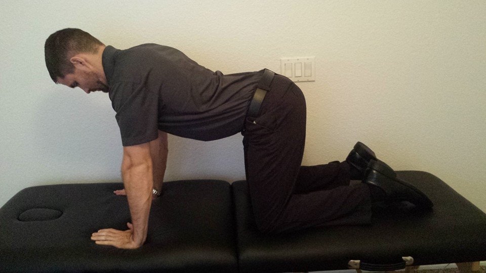
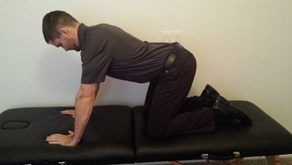
As many of you know, we recently launched a Home Exercise Portion to our website. They consist of many exercises that we prescribe and programs like VHI don't contain. I wanted to highlight one exercise today, the Quad Rock Back, because of all of uses for it. The exercise is a staple of the Shirley Sahrmann philosophy. While it is listed under the "Low Back" section, it is often prescribed for cervical, shoulder, and hip patients as well. We will break down how the exercise can be used for each region.

Picture
Picture
As described in the video, when this exercise is performed for cervical patients, often the head begins to extend or flex while rocking back. This is a result of abnormal movement and compensatory patterns in the cervical spine. For cervical spine patients, we encourage a chin tuck so that no neck movement occurs during the rocking.

There are many reasons why I like to give this exercise to shoulder and hip patients, one being joint compression. We are taught in our manual therapy classes that joint compression can be beneficial for healing; it also can help mobilize the posterior capsule. It also helps mimic the developmental patterns of weight bearing on the upper extremities, and thus is a critical part of rehabilitation. Additionally, the quad rock back exercise can be use to help normalize scapulohumeral movement patterns and avoid any compensatory activity. While performing the exercise, the movement can be completed by actively flexing the hips, instead of pushing away with the UE's. This allows the shoulder to move in a more normal pattern. Similarly, the exercise can help with unwanted hip musculature activity. By reversing the directions (instructing the patient to push with the hands and not actively flex the hips), the patient can decrease abnormal femoral head sliding and thus compete hip flexion less painfully.

The exercise can be used both for treating and assessing and low back dysfunctions, and not just because it is one of the more comfortable positions for low back pain. If you take your patient into quadruped and have them rock back, pay attention to the pelvis, hips, and low back. You may note a deviation to one side, suggesting increased muscle activity, stiffness, or movement patterns. Often you will note premature lumbar flexion compared to end-range hip flexion. This is secondary to all the sitting we do throughout the day - our lumbar spine becomes more flexible compared to the hip! By encouraging the patient to maintain a stable lumbar spine while rocking back (this can be done by placing a stick across the low back - if it falls off, we know there has been abnormal movement). This teaches our patients to isolate hip movement from back movement.

While I may not subscribe to all of Sahrmann's theories on movement impairment syndromes, especially in the acute phase, I do appreciate the focus she places on changing compensatory patterns with her exercises. Many of these patterns stem from abnormal postures or repetitive tasks that we perform throughout our daily lives. The rehab focuses on resisting any changes in movement that occur as a result. I have found that I get better results with incorporating repeated motions into my treatment, but I continued to use Sahrmann exercises to try and retrain movement afterwords.

-Chris
1 Comment

Sitting Wrecks Your Hip Action

8/29/2014

0 Comments

 
In the video below, Kelly Starrett discusses how sitting throughout the day places the hip joint in a poor position for normal daily movements. In the video he discusses how prolonged sitting places the psoas muscle in a short, contracted position. As an individual attempts to stand from this position, he/she hyper-extends the lumbar spine. Biomechanically, a tight psoas and shortened lumbar paraspinals draw the pelvis into an anterior pelvic tilt and the lumbar vertebrae into extension. Be aware: the chest may appear upright- giving the impression of natural movement,- but a closer look at the L-spine reveals movement beyond the neutral range.    

The problem with sitting exists beyond the chair as well. Normal standing posture requires little muscular action to maintain an upright position. In the presence of a hip flexor contracture due to prolonged sitting, forces are placed anteriorly across the hip requiring the hip extensors to counteract the force. Increased muscular action and metabolic cost creates the desire to sit, perpetuating the circumstance which initiated the hip flexion contracture (Neumann 2010). 
Quick facts regarding the hip joint:
-Normal hip flexion AROM: 120 degrees; extension AROM: 10-15 degrees
-Unmodified, ovoid ball and socket joint with 3 deg of freedom
-Capsular pattern (as defined by Cyriax): hip flexion > restricted hip ABDuction > restricted IR
-Closed packed position: Hip extension, ABDuction, IR
-Open packed position: 30 deg flexion, 30 deg abduction, 20 deg ER    
Quick facts regarding the psoas muscle:
-The psoas attaches proximally along the transverse process of T12 and all lumbar vertebrae
-The muscle attaches distally on the less trochanter of the femur
-The iliopsoas is a stong hip flexor and tilts the pelvis anteriorly as well
-If the pelvis is not stabilized the psoas will pull the lumbar spine into excessive lordosis
The Student Physical Therapist Advice: 
Engage the Transversus abdominus (TrA) prior to moving from sitting to standing. The TrA provides a posterior pelvic tilt to counteract the force of the psoas muscle. Additionally a posterior tilt does not allow the lumbar paraspinals to contract as readily. With proper cueing, the gluteus maximus can then be engaged and stress will be taken off the low back.  

Enjoy the video-  Jim 
References: Neumann, Donald. Kinesiology of the Musculoskeletal System: Foundations for Rehabilitation. 2nd edition. St. Louis, MO: Mosby Elsevier, 2010. 341-342. Print.
0 Comments

Hamstring Tightness or Neural Tension: How can you tell?

11/14/2013

6 Comments

 
Picture
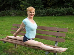
We are all guilty at one time or another of prescribing a hamstring stretch prematurely. You saw a patient bend forward to touch their toes and they abruptly stop short. This must be their hamstrings stopping their motion! Well unfortunately it is not that simple anymore. Modern literature tells us the reason they cannot touch their toes may be due to an array of possibilities: tight posterior chain muscles, adverse neural tensioning, or poor stability/motor control of the core musculature just to name a few. For the sake of this post we want to address hamstring tightness vs. adverse neural tension. 

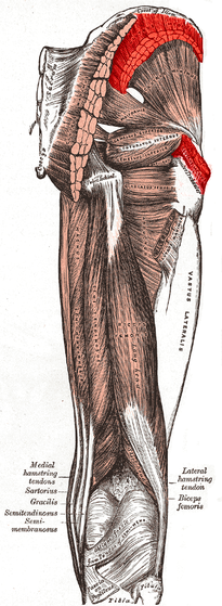
To begin this conversation, let's take that same patient above and place him/her in long sitting to reduce the effects of spinal loading. Now, postural demands are less than that in standing. We can focus specifically on muscle tightness vs. neural tension. The patient leans forward to touch their toes, but again nothing changes. We are no closer to finding our solution than we were in standing. Now, we bring the patient into supine, and the therapist performs a passive straight leg raise (PSLR). The patient cannot raise beyond 60 degrees of hip flexion. We still do not know, but we are getting close to the answer. Is it muscle tightness that is stopping this patient at 60 degrees or is the act of passively straightening the leg causing strain on the neural tissues. To answer this question, simply palpate the ASIS as you perform the PSLR. If you notice that hip flexion stops prior to movement at the ASIS, the answer is Neural Tension. If the ASIS begins to move prior to resistance, then hamstring tightness is the answer. 

Since the hamstrings attach to the Ischial Tuberosity, the body will naturally begin to posterior pelvic tilt once all the available range is taken up in the hamstring muscles. This posterior pelvic tilt will cause movement at the ASIS, letting you know the hamstring muscles have reached their end range. If no movement was noted at the ASIS, you can shift your hypothesis towards adverse neural tension. An additional component you can consider is the use of cervical flexion/extension to change the tension of the nerves and thus potentially alter SLR ROM.

Just like any examination test, it is important to cluster your findings with other tests and measures. For example, if you suspect adverse neural tension, perform a SLUMP test as well. Additionally, always check side to side symmetry. 

6 Comments

Gluteus Medius MMT: Was Kendall Wrong?

11/12/2013

3 Comments

 
Picture
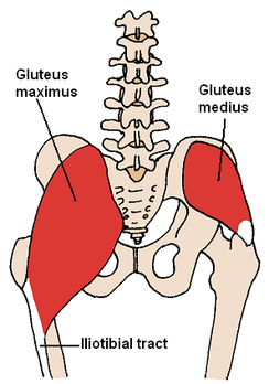
If you are familiar with Florence Kendall's method of Manual Muscle Testing (MMT), you are likely aware of the fact that she prefers to test muscle strength in a partially shortened position. When muscle length is in mid-range, it allows optimal cross-bridge linking, leading to a higher potential for force production. When muscles are over-extended or excessively shortened, the muscles lose that high frequency of cross-bridge linking. If you look at Kendall's MMT for the gluteus medius, you might see that she tries to emphasize the posterior fibers by having the patient on the side with the pelvis tilted forward and hip in extension, abduction, and lateral rotation. This is position completely shortens the muscle, thus potentially lower the validity of strength assessment. We have already discussed the use of evaluating hip abduction strength in a closed-chain position. Something else to consider is the fact that the gluteus medius is essential for pelvic stability throughout the entire stance phase of the gait cycle, not just mid-stance (where Kendall tests). Does it really matter if you have a level pelvis at midstance if you cannot keep your pelvis stable at heel strike? We have been presented another method for testing the gluteus medius in open-chain. Instead, place the lower extremity in hip abduction, lateral rotation, and flexion. The examiner then applies a flexion and adduction directed force. This position allows the muscle to not be completely shortened and tested in a functionally useful position. The position of flexion that is added can mimic heel strike in the gait cycle, making certain the muscle is strong enough to prevent pelvic drop as soon as stance begins.

3 Comments

Quick Gait Assessment

7/27/2013

0 Comments

 
Picture
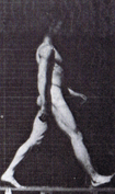
In the clinic, we often only have the length of the gym to analyze a patient's gait pattern. They patient may walk back and forth 2-3 times, giving you a total of 30 seconds to 1 minute to fully assess their pattern and recognize any deviations. 

In this post from the Physioblogger, he discusses his Quick Gait Assessment. His assessment includes looking at the feet, hips, and trunk. He starts at the feet and works cephalad. Beginning at the feet, he assesses heel to toe progression and the amount of pronation/supination. Next at the hips, he analyzes frontal plane motion and the amount of pelvic sway. Finally in the thoracic region, quick notes are made on the amount of arm swing and trunk stiffness.

Important points:
-No diagnosis is made during the gait assessment. Formulating hypotheses as to why a deviation is present is fine, but do not make definitive judgements as to why deviations are occurring. It is simply an observation. 
-Always remember a patient's injury, mechanism, lifestyle. What structures are involved? How can this injury/ their lifestyle impact the gait cycle?
-If possible, videotape your patients! Watch their gait patterns after they are gone. Take a few notes, perform the appropriate tests and measure, and act accordingly! 

Everyone has a different system they use for their quick gait assessment. Having a consistent method and practicing that method is important to your success as a therapist. Remember, we are movement analysis specialists. 

0 Comments

The Powers of Observation

7/2/2013

2 Comments

 
Picture
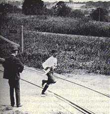
     If you feel like we having been referencing The Gait Guys a lot recently... it is because we have! They have put out a few great segments recently including this 2 part series on the Power of Observation. As clinical novices, we often do not have enough opportunities to see pathological gait patterns and conditions. As former students, almost of our lab "patients" were healthy classmates. While those experiences were rewarding, they were far from a real situation.   

     In this series, Shawn and Ivo dissect a triathlete's gait pattern. The patient presents with chronic low back pain, but has impairments throughout the entire lower chain. One interesting impairment they discuss is a "clenched fist on the L," which they attribute to flexor dominance and related to decreased arm swing and proprioceptive deficits. Their explanation: the proprioceptive system feeds the cerebellum. The cerebellum helps fire axial extensors. Because proprioception is limited in this athlete, the patient is naturally drawn into a flexor dominant position. Check out the entire post to gain more gems like this one! 

Because the gait guys use a Tumblr format, you may have to scroll down to June 25 and June 26. Enjoy!  

2 Comments

Muscle Activation During Various Angles of Clam Exercises

5/28/2013

5 Comments

 
A common exercise you may see in the clinic is sidelying clam shells. We had previously been hesitant to utilize this exercise ourselves, as we were taught that it preferentially activates the TFL over the Gluteus Medius. With weakness in the Gluteus Medius being a frequent culprit in individuals with knee, hip, and back pathologies, you can understand our avoidance of this exercise.

In JOSPT's recent issue, one of the studies reported on the muscle activation pattern in the hip for various angles of clam exercises. The authors looked at EMG activity of the Gluteus Maximus, Gluteus Medius, and TFL during sidelying clam shell exercises with the hip in 0, 30, and 60 degrees of flexion. Additionally, they studied the effect of complete sidelying and a 35 degree backward rotation (reclined) had on muscle activation. Interestingly, TFL activity was low throughout all positions, while Gluteus Maximus and Gluteus Medius were highest in complete sidelying with 60 degrees of hip flexion. This shows that, with proper positioning, sidelying clam shell exercises can be useful for strengthening the Gluteus Medius (along with the Gluteus Maximums).

That being said, there are two items that come to mind. As much as the Gluts are targeted with sidelying clam shells, do they have increased activation compared to pure sidelying hip abduction for the Gluteus Medius (which is the MMT position)? Obviously, we want to choose the exercises that focus on the muscle the most. This study looked at hip muscle activation in several exercises and found sidelying hip abduction to be the most specific to strengthening of the Gluteus Medius. Additionally, clam shells are sidelying. To what extent do we want to continue sidelying/supine exercises, when they are not as functional as more advanced, standing exercises? That's not to say that clam shells do not serve a purpose for patients at lower functional levels, but are we relying on lower level exercises too much?
Picture
5 Comments

The bio-future of joint replacement - TED Talk

4/27/2013

1 Comment

 
This is a good, quick 7-minute TED Talk given by the orthopaedic surgeon, Dr. Kevin Stone. He presents several interesting concepts and innovative strategies about the future of joint replacements. This might not be something you see in your clinical practice today, but as science continues to improve Dr. Stone's biological approach to joint replacements could be something you see on a more regular basis. Check it out!   
1 Comment

A Functional Method for Assessing the Glut Med

4/22/2013

0 Comments

 
Picture
We are all aware of the standard MMT for the gluteus medius in sidelying, but how well does this assessment transition to functional activities like walking? A sign we occasionally see is the Trendelenburg sign. This may consist of the uncompensated type (contralateral pelvic drop) or compensated type (ipsilateral trunk lean). Why not use Trendelenberg as a test as well? The Gait Guys had a recent post where they review this functional method of assessing gluteus medius strength. They include the various compensation methods to watch out for along with potential causes for the impairment. Check it out!

0 Comments

Trochanteric Bursitis or Hip Abductor Tear?

2/7/2013

2 Comments

 
    I recently had a patient with a history of being diagnosed with "trochanteric bursitis." While her therapy at the time was not for the hip, the patient often complained of pain superoposterior to the greater trochanter. This pain could be reproduced with ambulation and palpation. Having learned of bursitis being misdiagnosed before, I decided to delve into the topic. Upon further discussion, I learned she had the hip pain for approximately 5 years following being diagnosed with bursitis. The patient reported that no MRI was ordered for her hip. She also presented with a positive uncompensated trendelenburg during gait, pain with active hip abduction, pain-free passive hip abduction. Upon review of the literature, it appears there could potentially be a large incidence of hip abductor tears being missed in these patients. This article discusses how the findings of patients with hip abductor tears are shown to be similar to the signs of those diagnosed with bursitis. With the load our hip musculature withstands throughout a lifetime and the potential excessive load due to compensating for other gait deviations, perhaps we should be taking a second look at our elderly patients with hip pain that do not seem to be progressing.
Picture
2 Comments
<<Previous
    Picture
    Dr. Brian Schwabe's NEW Book in partner with PaleoHacks!
    Picture
    Learn residency-level content on our
    Insider Access pages
    Picture

    Picture

    We value quality PT education & CEU's.  Click the MedBridge logo below for TSPT savings!


    Archives

    July 2019
    June 2019
    May 2019
    March 2019
    February 2019
    January 2019
    December 2018
    November 2018
    October 2018
    September 2018
    August 2018
    July 2018
    June 2018
    May 2018
    April 2018
    March 2018
    February 2018
    January 2018
    December 2017
    November 2017
    October 2017
    September 2017
    August 2017
    July 2017
    June 2017
    May 2017
    April 2017
    March 2017
    February 2017
    January 2017
    December 2016
    November 2016
    October 2016
    September 2016
    August 2016
    July 2016
    June 2016
    May 2016
    April 2016
    March 2016
    February 2016
    January 2016
    December 2015
    November 2015
    October 2015
    September 2015
    August 2015
    July 2015
    June 2015
    May 2015
    April 2015
    March 2015
    February 2015
    January 2015
    December 2014
    November 2014
    October 2014
    September 2014
    August 2014
    July 2014
    June 2014
    May 2014
    April 2014
    March 2014
    February 2014
    January 2014
    December 2013
    November 2013
    October 2013
    September 2013
    August 2013
    July 2013
    June 2013
    May 2013
    April 2013
    March 2013
    February 2013
    January 2013
    December 2012
    November 2012
    October 2012
    September 2012
    August 2012

    Categories

    All
    Chest
    Core Muscle
    Elbow
    Foot
    Foot And Ankle
    Hip
    Knee
    Manual Therapy
    Modalities
    Motivation
    Neck
    Neural Tension
    Other
    Research
    Research Article
    Shoulder
    Sij
    Spine
    Sports
    Therapeutic Exercise


    RSS Feed

Home

Contact Us

Copyright © The Student Physical Therapist LLC 2022
Photos used under Creative Commons from dsearls, moo.review, liverpoolhls, matturick, lwpkommunikacio, Lake Clark NPS, Tobyotter, Charly Meunier, Vincent Albanese, CNBP, Zepfanman.com, aotaro, sportEX journals, jillccarlson, Yann07, US Department of Education, osseous, TheeErin, ajy591, Mark P / PIX41, Thomas Fisher Rare Book Library, UofT, teresatrimm, donnierayjones, NNelumba, Tobyotter, Frédéric de Villamil, Jordanhill School D&T Dept, danabooo, cadillacjr2002, ingridkreuz, RDECOM, U.S. Naval Forces Central Command/U.S. Fifth Fleet, Wonderlane, hectorir, Jeffrey, Chris Hunkeler, quillons, COD Newsroom, Alan Cleaver, CCFoodTravel.com, liverpoolhls, Dr.Farouk, Jeffrey, Sten Dueland, Håkan Dahlström, PPGWings, Parker Michael Knight, MLazarevski, dno1967b, Fimb, Jim Larrison, infomatique, U.S. Naval Forces Central Command/U.S. Fifth Fleet, Idhren, iwona_kellie, AnEternalGoldenBraid, quinn.anya, jlk.1, Villainette, mlsaero, roelandpype, Defence Images, CiscoANZ, istolethetv, Our Dream Photography (Personal), BraNewbs, Meg Stewart, phalinn, akeg, hectorir, philcampbell, MilitaryHealth, akeg, Army Medicine, Shar Ka, Subconsci Productions, Eric Kilby, Anonymous9000, quinn.anya, COD Newsroom, UMN Department of Plant Pathology, familymwr, all of olive., Keith Allison, JD Hancock, Synergy by Jasmine, roger_mommaerts, John-Morgan, a.drian, Instant Vantage, Beshef, markhillary, Vegar S Hansen Photography, DEC International, kaolin fire, smith_cl9, cinnamon_girl, jillccarlson, Extra Ketchup, brain_blogger, heyjoewhereyougoinwiththatguninyourhand, tv42, COMSALUD, Nicholas_T, Ano Lobb. @healthyrx, kaibara87, mlinksva, smbuckley23, sappsnap, Menage a Moi, brain_blogger, MaloMalverde, One Way Stock, ethet., ~ggvic~, MAClarke21, Keith Allison, pictures of money, mendolus shank, HotlantaVoyeur, Mr. T in DC, abbamouse, HystericalMark, Travis Hornung, jenny downing, shock264, Peter Mooney, Futurilla, sfslim, Emily Carlin, PFX Photo, ITU Pictures, TP studio, akunamatata, Magdalena Roeseler, osseous, smith_cl9, warrenski, Sh4rp_i, mrbichel, Renaud Camus, sicamp, GreenFlames09, infomatique, mark i geo, Volker Davids, TerryJohnston, Daniela Vladimirova, CJS*64, VarsityLife, Quentin Verwaerde, Annie Mole, US Army Africa, RLHyde, openBIT e. V., Vox Efx, fabola, Gerry Dincher, mlsaero, Infomastern, N4n0, Wonderlane, TheeErin, Nanagyei, gogogadgetscott, Pulmonary Pathology, PELeCON, Elvert Barnes, @RunRockPrincess, woodleywonderworks, Asbestos Bill, Joelk75, otisarchives4, jillccarlson, sportEX journals, The Wandering Angel, perpetualplum, V31S70, colecamp, jpalinsad360, JulyYu, Andres Rueda, Whenleavesfall, melloveschallah, Pink Sherbet Photography, canonim, BrianHenry ////|//, Leonard John Matthews, mikecogh, Zepfanman.com, Kevin M. Gill, Claire L. Evans, afunkydamsel, The Official CTBTO Photostream, cyOFdevelin fame, dok1, Pam loves pie, hoshi7, opensourceway
  • Home
  • About Us
  • Insider Access
    • About Insider Access
  • Online Courses
    • Hooper's Knee
    • Physical Therapist Entrepreneur Course
    • Physical Therapist Consultant Course
    • Orthopedic Management of the Cervical Spine
    • Return to Sport Essentials Course
  • Resources
    • Newsletter
    • Business Minded Sports Physio Podcast
    • Day in the Life of a Sports PT
    • Residency Corner
    • Special Tests >
      • Cervical Spine >
        • Alar Ligament Test
        • Bakody's Sign
        • Cervical Distraction Test
        • Cervical Rotation Lateral Flexion Test
        • Craniocervical Flexion Test (CCFT)
        • Deep Neck Flexor Endurance Test
        • Posterior-Anterior Segmental Mobility
        • Segmental Mobility
        • Sharp-Purser Test
        • Spurling's Maneuver
        • Transverse Ligament Test
        • ULNT - Median
        • ULNT - Radial
        • ULNT - Ulnar
        • Vertebral Artery Test
      • Thoracic Spine >
        • Adam's Forward Bend Test
        • Passive Neck Flexion Test
        • Thoracic Compression Test
        • Thoracic Distraction Test
        • Thoracic Foraminal Closure Test
      • Lumbar Spine/Sacroiliac Joint >
        • Active Sit-Up Test
        • Alternate Gillet Test
        • Crossed Straight Leg Raise Test
        • Extensor Endurance Test
        • FABER Test
        • Fortin's Sign
        • Gaenslen Test
        • Gillet Test
        • Gower's Sign
        • Lumbar Quadrant Test
        • POSH Test
        • Posteroanterior Mobility
        • Prone Knee Bend Test
        • Prone Instability Test
        • Resisted Abduction Test
        • Sacral Clearing Test
        • Seated Forward Flexion Test
        • SIJ Compression/Distraction Test
        • Slump Test
        • Sphinx Test
        • Spine Rotators & Multifidus Test
        • Squish Test
        • Standing Forward Flexion Test
        • Straight Leg Raise Test
        • Supine to Long Sit Test
      • Shoulder >
        • Active Compression Test
        • Anterior Apprehension
        • Biceps Load Test II
        • Drop Arm Sign
        • External Rotation Lag Sign
        • Hawkins-Kennedy Impingement Sign
        • Horizontal Adduction Test
        • Internal Rotation Lag Sign
        • Jobe Test
        • Ludington's Test
        • Neer Test
        • Painful Arc Sign
        • Pronated Load Test
        • Resisted Supination External Rotation Test
        • Speed's Test
        • Posterior Apprehension
        • Sulcus Sign
        • Thoracic Outlet Tests >
          • Adson's Test
          • Costoclavicular Brace
          • Hyperabduction Test
          • Roos (EAST)
        • Yergason's Test
      • Elbow >
        • Biceps Squeeze Test
        • Chair Sign
        • Cozen's Test
        • Elbow Extension Test
        • Medial Epicondylalgia Test
        • Mill's Test
        • Moving Valgus Stress Test
        • Push-up Sign
        • Ulnar Nerve Compression Test
        • Valgus Stress Test
        • Varus Stress Test
      • Wrist/Hand >
        • Allen's Test
        • Carpal Compression Test
        • Finkelstein Test
        • Phalen's Test
        • Reverse Phalen's Test
      • Hip >
        • Craig's Test
        • Dial Test
        • FABER Test
        • FAIR Test
        • Fitzgerald's Test
        • Hip Quadrant Test
        • Hop Test
        • Labral Anterior Impingement Test
        • Labral Posterior Impingement Test
        • Long-Axis Femoral Distraction Test
        • Noble Compression Test
        • Percussion Test
        • Sign of the Buttock
        • Trendelenburg Test
      • Knee >
        • Anterior Drawer Test
        • Dial Test (Tibial Rotation Test)
        • Joint Line Tenderness
        • Lachman Test
        • McMurray Test
        • Noble Compression Test
        • Pivot-Shift Test
        • Posterior Drawer Test
        • Posterior Sag Sign
        • Quad Active Test
        • Thessaly Test
        • Valgus Stress Test
        • Varus Stress Test
      • Foot/Ankle >
        • Anterior Drawer
        • Calf Squeeze Test
        • External Rotation Test
        • Fracture Screening Tests
        • Impingement Sign
        • Navicular Drop Test
        • Squeeze Test
        • Talar Tilt
        • Tarsal Tunnel Syndrome Test
        • Test for Interdigital Neuroma
        • Windlass Test