The Student​ Physical Therapist
  • Home
  • About Us
  • Insider Access
    • About Insider Access
  • Online Courses
    • Hooper's Knee
    • Physical Therapist Entrepreneur Course
    • Physical Therapist Consultant Course
    • Orthopedic Management of the Cervical Spine
    • Return to Sport Essentials Course
  • Resources
    • Newsletter
    • Business Minded Sports Physio Podcast
    • Day in the Life of a Sports PT
    • Residency Corner
    • Special Tests >
      • Cervical Spine >
        • Alar Ligament Test
        • Bakody's Sign
        • Cervical Distraction Test
        • Cervical Rotation Lateral Flexion Test
        • Craniocervical Flexion Test (CCFT)
        • Deep Neck Flexor Endurance Test
        • Posterior-Anterior Segmental Mobility
        • Segmental Mobility
        • Sharp-Purser Test
        • Spurling's Maneuver
        • Transverse Ligament Test
        • ULNT - Median
        • ULNT - Radial
        • ULNT - Ulnar
        • Vertebral Artery Test
      • Thoracic Spine >
        • Adam's Forward Bend Test
        • Passive Neck Flexion Test
        • Thoracic Compression Test
        • Thoracic Distraction Test
        • Thoracic Foraminal Closure Test
      • Lumbar Spine/Sacroiliac Joint >
        • Active Sit-Up Test
        • Alternate Gillet Test
        • Crossed Straight Leg Raise Test
        • Extensor Endurance Test
        • FABER Test
        • Fortin's Sign
        • Gaenslen Test
        • Gillet Test
        • Gower's Sign
        • Lumbar Quadrant Test
        • POSH Test
        • Posteroanterior Mobility
        • Prone Knee Bend Test
        • Prone Instability Test
        • Resisted Abduction Test
        • Sacral Clearing Test
        • Seated Forward Flexion Test
        • SIJ Compression/Distraction Test
        • Slump Test
        • Sphinx Test
        • Spine Rotators & Multifidus Test
        • Squish Test
        • Standing Forward Flexion Test
        • Straight Leg Raise Test
        • Supine to Long Sit Test
      • Shoulder >
        • Active Compression Test
        • Anterior Apprehension
        • Biceps Load Test II
        • Drop Arm Sign
        • External Rotation Lag Sign
        • Hawkins-Kennedy Impingement Sign
        • Horizontal Adduction Test
        • Internal Rotation Lag Sign
        • Jobe Test
        • Ludington's Test
        • Neer Test
        • Painful Arc Sign
        • Pronated Load Test
        • Resisted Supination External Rotation Test
        • Speed's Test
        • Posterior Apprehension
        • Sulcus Sign
        • Thoracic Outlet Tests >
          • Adson's Test
          • Costoclavicular Brace
          • Hyperabduction Test
          • Roos (EAST)
        • Yergason's Test
      • Elbow >
        • Biceps Squeeze Test
        • Chair Sign
        • Cozen's Test
        • Elbow Extension Test
        • Medial Epicondylalgia Test
        • Mill's Test
        • Moving Valgus Stress Test
        • Push-up Sign
        • Ulnar Nerve Compression Test
        • Valgus Stress Test
        • Varus Stress Test
      • Wrist/Hand >
        • Allen's Test
        • Carpal Compression Test
        • Finkelstein Test
        • Phalen's Test
        • Reverse Phalen's Test
      • Hip >
        • Craig's Test
        • Dial Test
        • FABER Test
        • FAIR Test
        • Fitzgerald's Test
        • Hip Quadrant Test
        • Hop Test
        • Labral Anterior Impingement Test
        • Labral Posterior Impingement Test
        • Long-Axis Femoral Distraction Test
        • Noble Compression Test
        • Percussion Test
        • Sign of the Buttock
        • Trendelenburg Test
      • Knee >
        • Anterior Drawer Test
        • Dial Test (Tibial Rotation Test)
        • Joint Line Tenderness
        • Lachman Test
        • McMurray Test
        • Noble Compression Test
        • Pivot-Shift Test
        • Posterior Drawer Test
        • Posterior Sag Sign
        • Quad Active Test
        • Thessaly Test
        • Valgus Stress Test
        • Varus Stress Test
      • Foot/Ankle >
        • Anterior Drawer
        • Calf Squeeze Test
        • External Rotation Test
        • Fracture Screening Tests
        • Impingement Sign
        • Navicular Drop Test
        • Squeeze Test
        • Talar Tilt
        • Tarsal Tunnel Syndrome Test
        • Test for Interdigital Neuroma
        • Windlass Test
Picture

How to Treat Neural Tension

10/28/2013

6 Comments

 
A couple weeks ago we reviewed the topic of Neurodynamics and along with how, when, and why we should test for neural tension. Due to the impact nerve irritation can have on your patient's symptoms, we thought we would review how to treat neural tension as well. Some of the techniques we will review include: nerve gliding/tensioning, nerve sliding, Active Release Therapy, and IASTM.

Often thought to be synonymous, in the next two paragraphs we will address the distinct differences between nerve gliding and nerve sliding exercises. 

Nerve gliding exercises try to separate the nerve from the surrounding structures by sliding the nerve via joint movements that lengthen the nerve bed (Coppieters & Butler, 2008). Lengthening the nerve bed also stretches the nerve. This can lead to increased neural tension and intraneural pressure. If this stretch is held statically, it can decrease blood flow to the nerves, thus leading to cell death. On the other hand, dynamic movements can assist in pumping out some of the edematous fluid that compresses the nerve. That being said, any time the nerve bed is elongated there is a chance for increasing symptoms. Due to the risk of increasing symptoms, these exercises are not as common as they once were.
Picture
A similar technique often confused with nerve gliding is nerve sliding. Nerve sliding works by elongating the nerve bed at one joint, while simultaneously shortening it at another (Coppieters & Butler, 2008). The reasoning is that the nerve can move without increasing strain. It was found that nerve sliding creates the largest nerve excursions with the least amount of strain. Nerve gliding can potentially create even larger nerve excursions at proximal joints but it creates significant strain. Due to the chance for symptom provocation, nerve gliding should only be considered in non-acute and non-surgical conditions. It is here that nerve sliding is the preferred intervention.

A recent study found that the sequencing of nerve tensioning/gliding at each joint was irrelevant when looking at net strain on the nerve (Boyd et al, 2013). However, it was also shown that variation in sequencing of joint movements altered where the nerve strain occurred first. This has potential clinical implications as we may be able to target specific locations if we know where the restrictions lie. The real-world applicability is unknown at this point as there have not been any studies performed in this area.

When comparing education to education + neural tissue management (both nerve sliding/gliding were used with cervical manual therapy), it was found that the intervention group had superior results compared to the control with no significant increased risk of exacerbations (Nee et al, 2012). Not only is it important to note the benefit of these nerve gliding/sliding exercises, but it brings up the point that we should also be looking at the spine. Other than some form of direct trauma, another source of nerve irritation can come from poor spinal mechanics that lead to neural irritation. Treating just the nerve may mean treating just the symptoms in some cases. It is essential to look at the spinal and restore normal mechanics if any abnormalities are found, especially because a manipulation may immediately show symptom relief as well.

There are two additional treatment techniques we wanted to mention. A case study we looked at utilized Active-Release Therapy (ART) for saphenous nerve entrapment (Settergren, 2012). In general, ART involves a technique where the clinician applies a force to the restricted area while the patient actively moves to "release" the adhesion. The technique often causes significant pain during the maneuver but is followed by increased mobility and decreased pain. This method may not be as useful to most clinicians as it involves extensive training to correctly perform and the research is limited in the area. Another technique that we often perform and have had success with is Instrument-Assisted Soft Tissue Mobilization (IASTM). While we have personally seen immediate effects on pain and neural symptoms with this, again the research is limited in the area.
References:
Boyd BS, Topp KS, & Coppieters MW. (2013). Impact of Movement Sequencing on Sciatic and Tibial Nerve Strain and Excursion During the Straight Leg Raise Test in Embalmed Cadavers. JOSPT 2013 43(6):398-403.

Coppieters MW & Butler D. (2008). Do "sliders" slide and "tensioners" tension? An Analysis of Neurodynamic Techniques and Considerations Regarding Their Application. Manual Therapy 2008 13(3): 213-221. Web. 26 October 2013.

Nee RJ, Vicenzino B, Jull GA, Cleland JA, and Coppieters MW. (2012). Neural Tissue Management Provides Immediate Clinically Relevant Benefits Without Harmful Effects For Patients With Nerve-Related Neck and Arm Pain: A Randomised Trial. Journal of Physiotherapy 58 2012. Web. 26 October 2013.

Settergren R. (2012). Conservative Management of a Saphenous Nerve Entrapment in a Female Ulra-Marathon Runner. J Bodyw Mov Ther. 2013 Jul;17(3):297-301. Web. 26 October 2013.
6 Comments

Best Special Tests for Subacromial Impingement

10/24/2013

0 Comments

 
Picture
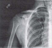
Subacromial Impingement Syndrome (SAIS) is reported to be the most frequent cause of shoulder pain in an OP physical therapy clinic. Despite the high prevalence, physical therapists still struggle to appropriately diagnose the syndrome. The gold standard for diagnosing SAIS in arthroscopic surgery. Since we do not have access to this tool everyday, we must reply on our patient examination skills. A study by Michener et al, Reliability and Diagnostic Accuracy of 5 Physical Examination Tests and Combination of Tests for Subacromial Impingement, assessed 5 special tests commonly used to help rule-in SAIS. The 5 tests were Neers, Hawkins-Kennedy, Painful Arc, Empty Can (Jobe), and External Rotation Resistance Test. Specifically this article wanted to assess the interrator reliability of the tests, diagnostic accuracy of each test, and finally if clustering the tests would confirm or rule-out SAIS.


     The results of the study were surprising. Moderate to Substantial strength of agreement between raters was found for the empty can test, the painful arc sign, and external rotation resistance test. Surprisingly Hawkins Kennedy had the lowest kappa value at .39. We found this surprising because Hawkins-Kennedy is graded as painful or not painful. There seems to be little room for subjectivity, yet it received the lowest reliability among raters of all the tests. The difference likely lies in how the test is performed and thus perceived. It is easy to forget to not horizontally adduct the shoulder sufficiently or to ignore the patient's compensation of elevating the tested shoulder during IR as a means of avoiding/minimizing the pain provocation. Something we must always be wary of in studies of manual techniques is accepting the fact that all examiners perform/analyze movements the same.


     When looking at the diagnostic accuracy of each test individually, the External rotation resistance test had the highest positive likelihood ratio (LR) of 4.39, empty can had the second highest with 3.9, and the painful arc sign came in third at positive LR 2.25. Finally, the article found that when clustering the tests a "combination of any 3 positive tests out of the 5 have the best ability to confirm SAIS, with small to moderate shifts in the pretest to posttest probability."    

      While this article provides interesting and useful clinical information, the results are different from other studies in the literature. A separate article on SAIS by Park et al found that the clustering Hawkins-Kennedy, Infraspinatus Muscle Test, and the Painful Arc Sign yielded a high +LR (10.56) for ruling-in SAIS. So what cluster should you use? Personally, we would use both. The etiology of SAIS is multifactorial. Several structures have the potential to be pain generators and the presentation of SAIS will vary based on posture, scapulohumeral rhythm, accessory joint mechanics, and more. The Michener article was quick to point out the importance of a thorough subjective history to help aide in the diagnostic process.  

Reference: Michener LA, Walsworth MK, Doukas WC, Murphy KP. Reliability and diagnostic accuracy of 5 physical examination 
     tests and combination of tests for subacromial impingement. Arch Phys Med Rehabil. 2009 Nov;90(11):1898-903.
Park HB, Yokota A, Gill HS, El Rassi G, McFarland EG. Diagnostic accuracy of clinical tests for the different degrees 
     of subacromial impingement syndrome. J Bone Joint Surg Am. 2005 Jul;87(7):1446-55.

0 Comments

Why We Must Utilize More Than Pain Provocation Tests for SIJ Dysfunction

10/21/2013

0 Comments

 
Picture
We are once again back on the topic of SIJ Dysfunction. A couple months ago, we had a pretty controversial post about non-painful sacroiliac joint dysfunction. A large part of our discussion was on the basis of how limitations in SIJ mobility could alter the normal stresses in adjacent joints. While we still believe this is an important aspect, in the past few weeks we have become aware of yet another reason for regularly assessing the sacroiliac joint.

Something that is often lost in the effects of SIJ dysfunction is impact on muscle alignment. Any time there is a deficiency in the alignment of the joint surfaces, the origin or insertion of the muscles are altered, thus changing the moment arm of the muscle as it crosses its respective joint. These changes may seem small and insignificant, but they can result in both muscle pain and weakness. The significance of a muscle alignment issue is that these patients will likely not have many positive pain provocation tests, because it is not the joint that is painful - it is the muscle (or structures near the muscle). Therefore, when assessing the sacroiliac joint, be sure to assess the symmetry and depth of SIJ structures such as the PSIS, sacral sulci, sacral ILAs, ischial tuberosities, and more. In the past few weeks, we have had multiple patients with pain or weakness that was eliminated or minimized simply by doing a sacral mobilization or other manual technique to correct the asymmetry. These patient's had negative pain provocation testing but noticeable bony landmark asymmetries. We are aware of the fact that SI mobility tests are unreliable according to several research studies, but we again argue that they should not be forgotten. This brings up the importance of testing and re-testing after a treatment. A muscle that appeared weak initially in your eval may be normal after correction. If you can show immediate changes in pain and strength from a treatment directed at the SIJ, not only will it potentially alter your plan of care, but it also builds your patients' confidence in you as a clinician! 

0 Comments

Neurodynamics

10/18/2013

2 Comments

 
Picture
Neurodynamics, Neural flossing, Nerve glides...what do these terms mean exactly? The concept of placing a mechanical stress on the nervous system is abstract for many students. While many people learn the common "tensioning tests" in a school lab or at a con-ed course, the knowledge of when to perform these tests AND how to interpret the tests is lost by many individuals. 
    
Clinicians often use neural tensioning tests to determine if a patient has adverse neural tension. If a part of the nervous system becomes inflamed, entrapped, or damaged, the patient will present with signs and symptoms of neural origin. Examples of diagnoses seen with adverse neural tension include carpal tunnel syndrome, cervical radiculopathy, and cubital tunnel syndrome.  By performing tensioning tests, the clinician can determine if the origin (or a component) of pain (or other symptoms) is neural or non-neural. Pain is not your only "positive" finding in the test. Pulling and stretching are just as significant. Basically, if you develop pain/pulling/stretching in an area when tensioning the nerve and it is modified by cervical movement, you need to consider nerve components. A study published in JOSPT in 2012 by Nee et al gives "probable" criteria in regards to diagnosing peripheral neuropathic pain: 1. symptoms fit a nerve related pattern 2. history of symptoms correlates with a nerve related problem. 3. a clinical neuro examination or imaging rule-in or confirm a neurological injury. 

If you suspect adverse neural tension to be contributing to your patient's symptoms, perform a neurodynamic test to help confirm your hypothesis. For example, if you suspect a patient has carpal tunnel syndrome, perform the Median Nerve ULTT first and foremost because that is the nerve most directly involved. When performing the tests, be cautious of false positive results. Many asymptomatic individuals will have adverse neural tension so you must ask yourself if the symptom response is relevant. A general 3-part rule I follow is A. does the + test reproduce the symptoms the patient came to see you for, B. is there a side to side difference, and c. Does the neural response change by moving a distant component. If these 3 components are true, adverse neural tension is likely contributing to your patient's pain. Now this is a controversial topic. Some people follow this 3-step rule. Others look for any findings of neural tension at all (not just pain and not just at the location of chief complaint) as a possible component to the pathology. Why? Because it is not normal.

There are neural tension for both the upper and lower extremities. Many people have heard of the SLUMP, Straight Leg Raise Tests, and ULTTs. You can check out how to perform the ULTT tests below:
1) Median Nerve ULTT
2) Radial Nerve ULTT
3) Ulnar Nerve ULTT

Why is it that we have neural tension and what do we do about the positive findings? Remember, neural tension is not normal, just like hypertonicity is not normal. Trauma (micro or macro) often is culprit. That trauma can be local to the nerve or as a result of abnormal spinal motion. For example, if you have excessive lumbar mobility on one side compared to another, eventually this stress can damage the nerves, leading to pain down the chain. Sound ridiculous? We have seen where a patient had greater lumbar extension R compared to L and had positive SLUMP testing with calf pain. Following a manipulation to the hypomobile side of the lumbar spine, the pain was significantly reduced. Nerve flossing is important for improving the mobility of neural tissue, but we must not forget to correct the original factor that led to the neural tension. In an upcoming post, we will review various methods of how to treat neural tensioning in general along with some other information.

The tests above with help you determine if adverse neural tension is present. The videos below by Chris Johnson will guide your intervention selection.
Commonly used Neurodynamic Interventions by Chris Johnson


As stated early, there are many false positive with neural tensioning tests. It is important that you be consistent and precise with your tension tests to get accurate results. Also, be sure to clarify your patient's responses and watch for compensations in the test positions. A slight variation can completely lose the pathological nerve tension.

References:
Nee RJ, et al. (2012). The Validity of Upper-Limb Neurodynamic Testing for detecting Peripheral Neuropathic 
     Pain. JOSPT. 2012 May; 42(5): 413-424. Web. 13 October 2013.  

2 Comments

10 Tips for Primary Care Residents/ Fellows and Medical Students

10/10/2013

2 Comments

 
Picture
Have you ever found yourself talking with Medical students or even doctors and wanted to give them some advice regarding referrals, treatments, or pathology in general. Well we definitely have been in this situation. Many medical school programs receive no formal education in Kinesiology or Biomechanics. As therapists, our scope on knowledge on regional interdependence in much greater. Specialists in Sports and Ortho Rehab (SSOR) recently came out with a post regarding 10 tips on how to educate primary care students/ residents/ fellows. 
The 10 tips are listed below, but check out the full post for in depth reading on the topic. 

1. Shoulder pain can be disguised as neck pain (and vice versa).
2a. Not all tendinopathies are the same.
2b. Eccentric exercises can be a promising intervention for certain tendinopathies.
3. Signs and symptoms of mensical tears. How to spot them during your physical exam!  
4. For people with general knee pain, address the hip abductors and external rotators.
5. Headaches can be treated by physical therapists.
6. Sciatica can be managed by postural education, core strengthening, nerve glides, and hip strengthening.
7. Carpal Tunnel isn't always just in the wrist.
8. In acute ankle sprains, give your patients an assistive device before having them walk around with a limp. 
9. In patients with low back pain or SI pain, the problem may not be in the back.
10. Modalities are misused and abused, and frankly, there is little evidence supporting them. 

These points may all seem very obvious to physical therapists, but they may not be so clear to everyone else. For example, how many times have you had a referral for low back pain when the cause of the dysfunction was not the low back at all. This is a good top 10 list, but it is not a complete list. Can you think of other beneficial tips on how to educate medical students/residents? 

2 Comments

Repeated Motions and Directional Preference

10/7/2013

0 Comments

 
When most people think of McKenzie method, they think of extension based exercises and herniated disks. That is but the tip of the iceberg. A true understanding of the system that McKenzie offers can provide tools to help clinicians both diagnose and treat various conditions in the spine and extremities. In a recent post, Dr. E from The Manual Therapist reviews exactly what directional preference is and why we sometimes need to test/treat with sustained or repeated motions. Directional preference refers to motion in any body part that needs to be sustained or repeated to end range in order to improve ROM, pain, DTRs, strength, and/or function. Dr. E provides common treatment methods throughout the spine in this post. He doesn't discuss loading in the extremity as much here, but that isn't the point. The purpose of this article is to raise the importance of determining if/where the directional preference lies, and how to treat it. Definitely worth checking out.
Picture
0 Comments
    Picture
    Dr. Brian Schwabe's NEW Book in partner with PaleoHacks!
    Picture
    Learn residency-level content on our
    Insider Access pages
    Picture

    Picture

    We value quality PT education & CEU's.  Click the MedBridge logo below for TSPT savings!


    Archives

    July 2019
    June 2019
    May 2019
    March 2019
    February 2019
    January 2019
    December 2018
    November 2018
    October 2018
    September 2018
    August 2018
    July 2018
    June 2018
    May 2018
    April 2018
    March 2018
    February 2018
    January 2018
    December 2017
    November 2017
    October 2017
    September 2017
    August 2017
    July 2017
    June 2017
    May 2017
    April 2017
    March 2017
    February 2017
    January 2017
    December 2016
    November 2016
    October 2016
    September 2016
    August 2016
    July 2016
    June 2016
    May 2016
    April 2016
    March 2016
    February 2016
    January 2016
    December 2015
    November 2015
    October 2015
    September 2015
    August 2015
    July 2015
    June 2015
    May 2015
    April 2015
    March 2015
    February 2015
    January 2015
    December 2014
    November 2014
    October 2014
    September 2014
    August 2014
    July 2014
    June 2014
    May 2014
    April 2014
    March 2014
    February 2014
    January 2014
    December 2013
    November 2013
    October 2013
    September 2013
    August 2013
    July 2013
    June 2013
    May 2013
    April 2013
    March 2013
    February 2013
    January 2013
    December 2012
    November 2012
    October 2012
    September 2012
    August 2012

    Categories

    All
    Chest
    Core Muscle
    Elbow
    Foot
    Foot And Ankle
    Hip
    Knee
    Manual Therapy
    Modalities
    Motivation
    Neck
    Neural Tension
    Other
    Research
    Research Article
    Shoulder
    Sij
    Spine
    Sports
    Therapeutic Exercise


    RSS Feed

Home

Contact Us

Copyright © The Student Physical Therapist LLC 2022
Photos used under Creative Commons from dsearls, moo.review, liverpoolhls, matturick, lwpkommunikacio, Lake Clark NPS, Tobyotter, Charly Meunier, Vincent Albanese, CNBP, Zepfanman.com, aotaro, sportEX journals, jillccarlson, Yann07, US Department of Education, osseous, TheeErin, ajy591, Mark P / PIX41, Thomas Fisher Rare Book Library, UofT, teresatrimm, donnierayjones, NNelumba, Tobyotter, Frédéric de Villamil, Jordanhill School D&T Dept, danabooo, cadillacjr2002, ingridkreuz, RDECOM, U.S. Naval Forces Central Command/U.S. Fifth Fleet, Wonderlane, hectorir, Jeffrey, Chris Hunkeler, quillons, COD Newsroom, Alan Cleaver, CCFoodTravel.com, liverpoolhls, Dr.Farouk, Jeffrey, Sten Dueland, Håkan Dahlström, PPGWings, Parker Michael Knight, MLazarevski, dno1967b, Fimb, Jim Larrison, infomatique, U.S. Naval Forces Central Command/U.S. Fifth Fleet, Idhren, iwona_kellie, AnEternalGoldenBraid, quinn.anya, jlk.1, Villainette, mlsaero, roelandpype, Defence Images, CiscoANZ, istolethetv, Our Dream Photography (Personal), BraNewbs, Meg Stewart, phalinn, akeg, hectorir, philcampbell, MilitaryHealth, akeg, Army Medicine, Shar Ka, Subconsci Productions, Eric Kilby, Anonymous9000, quinn.anya, COD Newsroom, UMN Department of Plant Pathology, familymwr, all of olive., Keith Allison, JD Hancock, Synergy by Jasmine, roger_mommaerts, John-Morgan, a.drian, Instant Vantage, Beshef, markhillary, Vegar S Hansen Photography, DEC International, kaolin fire, smith_cl9, cinnamon_girl, jillccarlson, Extra Ketchup, brain_blogger, heyjoewhereyougoinwiththatguninyourhand, tv42, COMSALUD, Nicholas_T, Ano Lobb. @healthyrx, kaibara87, mlinksva, smbuckley23, sappsnap, Menage a Moi, brain_blogger, MaloMalverde, One Way Stock, ethet., ~ggvic~, MAClarke21, Keith Allison, pictures of money, mendolus shank, HotlantaVoyeur, Mr. T in DC, abbamouse, HystericalMark, Travis Hornung, jenny downing, shock264, Peter Mooney, Futurilla, sfslim, Emily Carlin, PFX Photo, ITU Pictures, TP studio, akunamatata, Magdalena Roeseler, osseous, smith_cl9, warrenski, Sh4rp_i, mrbichel, Renaud Camus, sicamp, GreenFlames09, infomatique, mark i geo, Volker Davids, TerryJohnston, Daniela Vladimirova, CJS*64, VarsityLife, Quentin Verwaerde, Annie Mole, US Army Africa, RLHyde, openBIT e. V., Vox Efx, fabola, Gerry Dincher, mlsaero, Infomastern, N4n0, Wonderlane, TheeErin, Nanagyei, gogogadgetscott, Pulmonary Pathology, PELeCON, Elvert Barnes, @RunRockPrincess, woodleywonderworks, Asbestos Bill, Joelk75, otisarchives4, jillccarlson, sportEX journals, The Wandering Angel, perpetualplum, V31S70, colecamp, jpalinsad360, JulyYu, Andres Rueda, Whenleavesfall, melloveschallah, Pink Sherbet Photography, canonim, BrianHenry ////|//, Leonard John Matthews, mikecogh, Zepfanman.com, Kevin M. Gill, Claire L. Evans, afunkydamsel, The Official CTBTO Photostream, cyOFdevelin fame, dok1, Pam loves pie, hoshi7, opensourceway
  • Home
  • About Us
  • Insider Access
    • About Insider Access
  • Online Courses
    • Hooper's Knee
    • Physical Therapist Entrepreneur Course
    • Physical Therapist Consultant Course
    • Orthopedic Management of the Cervical Spine
    • Return to Sport Essentials Course
  • Resources
    • Newsletter
    • Business Minded Sports Physio Podcast
    • Day in the Life of a Sports PT
    • Residency Corner
    • Special Tests >
      • Cervical Spine >
        • Alar Ligament Test
        • Bakody's Sign
        • Cervical Distraction Test
        • Cervical Rotation Lateral Flexion Test
        • Craniocervical Flexion Test (CCFT)
        • Deep Neck Flexor Endurance Test
        • Posterior-Anterior Segmental Mobility
        • Segmental Mobility
        • Sharp-Purser Test
        • Spurling's Maneuver
        • Transverse Ligament Test
        • ULNT - Median
        • ULNT - Radial
        • ULNT - Ulnar
        • Vertebral Artery Test
      • Thoracic Spine >
        • Adam's Forward Bend Test
        • Passive Neck Flexion Test
        • Thoracic Compression Test
        • Thoracic Distraction Test
        • Thoracic Foraminal Closure Test
      • Lumbar Spine/Sacroiliac Joint >
        • Active Sit-Up Test
        • Alternate Gillet Test
        • Crossed Straight Leg Raise Test
        • Extensor Endurance Test
        • FABER Test
        • Fortin's Sign
        • Gaenslen Test
        • Gillet Test
        • Gower's Sign
        • Lumbar Quadrant Test
        • POSH Test
        • Posteroanterior Mobility
        • Prone Knee Bend Test
        • Prone Instability Test
        • Resisted Abduction Test
        • Sacral Clearing Test
        • Seated Forward Flexion Test
        • SIJ Compression/Distraction Test
        • Slump Test
        • Sphinx Test
        • Spine Rotators & Multifidus Test
        • Squish Test
        • Standing Forward Flexion Test
        • Straight Leg Raise Test
        • Supine to Long Sit Test
      • Shoulder >
        • Active Compression Test
        • Anterior Apprehension
        • Biceps Load Test II
        • Drop Arm Sign
        • External Rotation Lag Sign
        • Hawkins-Kennedy Impingement Sign
        • Horizontal Adduction Test
        • Internal Rotation Lag Sign
        • Jobe Test
        • Ludington's Test
        • Neer Test
        • Painful Arc Sign
        • Pronated Load Test
        • Resisted Supination External Rotation Test
        • Speed's Test
        • Posterior Apprehension
        • Sulcus Sign
        • Thoracic Outlet Tests >
          • Adson's Test
          • Costoclavicular Brace
          • Hyperabduction Test
          • Roos (EAST)
        • Yergason's Test
      • Elbow >
        • Biceps Squeeze Test
        • Chair Sign
        • Cozen's Test
        • Elbow Extension Test
        • Medial Epicondylalgia Test
        • Mill's Test
        • Moving Valgus Stress Test
        • Push-up Sign
        • Ulnar Nerve Compression Test
        • Valgus Stress Test
        • Varus Stress Test
      • Wrist/Hand >
        • Allen's Test
        • Carpal Compression Test
        • Finkelstein Test
        • Phalen's Test
        • Reverse Phalen's Test
      • Hip >
        • Craig's Test
        • Dial Test
        • FABER Test
        • FAIR Test
        • Fitzgerald's Test
        • Hip Quadrant Test
        • Hop Test
        • Labral Anterior Impingement Test
        • Labral Posterior Impingement Test
        • Long-Axis Femoral Distraction Test
        • Noble Compression Test
        • Percussion Test
        • Sign of the Buttock
        • Trendelenburg Test
      • Knee >
        • Anterior Drawer Test
        • Dial Test (Tibial Rotation Test)
        • Joint Line Tenderness
        • Lachman Test
        • McMurray Test
        • Noble Compression Test
        • Pivot-Shift Test
        • Posterior Drawer Test
        • Posterior Sag Sign
        • Quad Active Test
        • Thessaly Test
        • Valgus Stress Test
        • Varus Stress Test
      • Foot/Ankle >
        • Anterior Drawer
        • Calf Squeeze Test
        • External Rotation Test
        • Fracture Screening Tests
        • Impingement Sign
        • Navicular Drop Test
        • Squeeze Test
        • Talar Tilt
        • Tarsal Tunnel Syndrome Test
        • Test for Interdigital Neuroma
        • Windlass Test