The Student​ Physical Therapist
  • Home
  • About Us
  • Insider Access
    • About Insider Access
  • Online Courses
    • Hooper's Knee
    • Physical Therapist Entrepreneur Course
    • Physical Therapist Consultant Course
    • Orthopedic Management of the Cervical Spine
    • Return to Sport Essentials Course
  • Resources
    • Newsletter
    • Business Minded Sports Physio Podcast
    • Day in the Life of a Sports PT
    • Residency Corner
    • Special Tests >
      • Cervical Spine >
        • Alar Ligament Test
        • Bakody's Sign
        • Cervical Distraction Test
        • Cervical Rotation Lateral Flexion Test
        • Craniocervical Flexion Test (CCFT)
        • Deep Neck Flexor Endurance Test
        • Posterior-Anterior Segmental Mobility
        • Segmental Mobility
        • Sharp-Purser Test
        • Spurling's Maneuver
        • Transverse Ligament Test
        • ULNT - Median
        • ULNT - Radial
        • ULNT - Ulnar
        • Vertebral Artery Test
      • Thoracic Spine >
        • Adam's Forward Bend Test
        • Passive Neck Flexion Test
        • Thoracic Compression Test
        • Thoracic Distraction Test
        • Thoracic Foraminal Closure Test
      • Lumbar Spine/Sacroiliac Joint >
        • Active Sit-Up Test
        • Alternate Gillet Test
        • Crossed Straight Leg Raise Test
        • Extensor Endurance Test
        • FABER Test
        • Fortin's Sign
        • Gaenslen Test
        • Gillet Test
        • Gower's Sign
        • Lumbar Quadrant Test
        • POSH Test
        • Posteroanterior Mobility
        • Prone Knee Bend Test
        • Prone Instability Test
        • Resisted Abduction Test
        • Sacral Clearing Test
        • Seated Forward Flexion Test
        • SIJ Compression/Distraction Test
        • Slump Test
        • Sphinx Test
        • Spine Rotators & Multifidus Test
        • Squish Test
        • Standing Forward Flexion Test
        • Straight Leg Raise Test
        • Supine to Long Sit Test
      • Shoulder >
        • Active Compression Test
        • Anterior Apprehension
        • Biceps Load Test II
        • Drop Arm Sign
        • External Rotation Lag Sign
        • Hawkins-Kennedy Impingement Sign
        • Horizontal Adduction Test
        • Internal Rotation Lag Sign
        • Jobe Test
        • Ludington's Test
        • Neer Test
        • Painful Arc Sign
        • Pronated Load Test
        • Resisted Supination External Rotation Test
        • Speed's Test
        • Posterior Apprehension
        • Sulcus Sign
        • Thoracic Outlet Tests >
          • Adson's Test
          • Costoclavicular Brace
          • Hyperabduction Test
          • Roos (EAST)
        • Yergason's Test
      • Elbow >
        • Biceps Squeeze Test
        • Chair Sign
        • Cozen's Test
        • Elbow Extension Test
        • Medial Epicondylalgia Test
        • Mill's Test
        • Moving Valgus Stress Test
        • Push-up Sign
        • Ulnar Nerve Compression Test
        • Valgus Stress Test
        • Varus Stress Test
      • Wrist/Hand >
        • Allen's Test
        • Carpal Compression Test
        • Finkelstein Test
        • Phalen's Test
        • Reverse Phalen's Test
      • Hip >
        • Craig's Test
        • Dial Test
        • FABER Test
        • FAIR Test
        • Fitzgerald's Test
        • Hip Quadrant Test
        • Hop Test
        • Labral Anterior Impingement Test
        • Labral Posterior Impingement Test
        • Long-Axis Femoral Distraction Test
        • Noble Compression Test
        • Percussion Test
        • Sign of the Buttock
        • Trendelenburg Test
      • Knee >
        • Anterior Drawer Test
        • Dial Test (Tibial Rotation Test)
        • Joint Line Tenderness
        • Lachman Test
        • McMurray Test
        • Noble Compression Test
        • Pivot-Shift Test
        • Posterior Drawer Test
        • Posterior Sag Sign
        • Quad Active Test
        • Thessaly Test
        • Valgus Stress Test
        • Varus Stress Test
      • Foot/Ankle >
        • Anterior Drawer
        • Calf Squeeze Test
        • External Rotation Test
        • Fracture Screening Tests
        • Impingement Sign
        • Navicular Drop Test
        • Squeeze Test
        • Talar Tilt
        • Tarsal Tunnel Syndrome Test
        • Test for Interdigital Neuroma
        • Windlass Test
Picture

Plantar Fasciopathy 

10/13/2012

1 Comment

 
Picture
     It has been estimated that Plantar Fasciitis occurs in approximately 2 million people and can account for between 8% and 15% of all foot pain complaints. While the term "-itis" is often associated with inflammation, there is growing evidence that there might not be an inflammatory state, but rather a degenerative process occurring in the plantar fascia. Because of this growing belief, authors are saying a more appropriate term would be "plantar fasciopathy" or "plantar heel pain."  
    
    Plantar heel pain is best described as a sharp pain in the patient's rear foot that is worse in the morning (usually the first step out of bed) and at the beginning of a weight bearing activities. The pain typically lessens with continued activity, but often increases toward the end of the day. Individuals most susceptible to developing heel pain are middle-aged women, obese individuals, athletes, and runners. Clinically, you will see excessive pronation and a depressed longitudinal arch in many of these clients. Some extrinsic factors contributing to the pathology include training surfaces, shoe wear, and poor training methods. Understanding the anatomy makes it clear why this population is at an increased risk. The plantar fascia runs from the medial tuberosity of the calcaneus and inserts into the metatarsophalangeal joints, the proximal phalanges, and the flexor tendon sheaths. The fascia is responsible for supporting the longitudinal arch of the foot and assisting in dynamic shock absorption. The attachment of the plantar fascia to the medial calcaneal tuberosity explains why patients often experience pain upon palpation of that area.
   
    Diagnosis of plantar fasciopathy is often made on a clinical basis. Due to degenerative changes and tendon thickening, the diagnosis may be made with an ultrasound as well.  Current treatment methods include rest, modalities, stretching, strengthening, manual therapy, splinting, orthotics, surgery, and more. New research is constantly being published due to the high incidence of the injury.  This review will take an in depth look at several of the available treatment techniques for plantar fasciopathy. Many of the studies we looked at included strengthening and stretching in the treatment plans along with some other intervention. Improvement was often shown in both groups, but we were unable to find any studies that specifically looked at one type of strengthening exercise compared to another. Some of the most common barefoot exercises seen in the clinic include towel scrunching and picking up marbles. Due to the biomechanical theory of the plantar fascia aiding in the support of the medial arch, it would seem logical to include strengthening of the posterior tibialis in rehabilitation. The posterior tibialis is the prime muscle for raising the medial longitudinal arch and can take stress off the plantar fascia. As noted in our previous posts, the exercise to most effectively activate the posterior tibialis is resisted forefoot adduction.

      A 2011 randomized control trial we looked at compared the effectiveness of a self-directed stretching program to a stretching program plus trigger point release therapy. The stretching only group was instructed to perform a gastroc stretch, soleus stretch, and seated plantar fascia-specific stretch. The trigger point group performed the same stretching, but also received trigger point release to the gastroc and soleus muscles. While both groups improved, the trigger point therapy group reported higher physical function, less bodily pain, and increased pressure pain thresholds for short term outcomes.   
Picture
     Another study we reviewed compared a new calcaneal taping technique versus a sham taping group, a stretching group, and a no treatment group. The calcaneal taping technique inverted the heel to raise the medial longitudinal arch. A first piece of tape pulled the calcaneus medially. Pieces 2 and 3 followed the same pattern, overlapping 1/3 of each prior piece of tape. Piece 4 wrapped around the heel lateral to medial, supporting the arch and anchoring pieces 1-3. This 4 piece technique was considered quick and cost effective. This calcaneal taping intervention resulted in significantly greater reductions in pain compared to the sham taping, stretching, and no treatment groups.  Additionally, a study comparing Medial Arch Support to Low-Dye Taping found that both groups had improvements in pain, but the Medial Arch Support had significantly greater improvements. These interventions should be considered for short-term relief, so that the patient can be pain-free for more intense therapy or activities.
    
    A third intervention we reviewed assessed the effects of low level laser therapy in the treatment of plantar fasciitis. Laser treatments were given 3 times per week for 4 weeks with a 30mW . 83 um continuous-wave IR diode laser. The goal of the laser therapy was to alter cellular metabolism, protein synthesis, and create an immune response. The conclusions of randomized controlled evaluation found that low level laser therapy was not beneficial in the treatment of plantar heel pain. A study we looked at compared the effects of stretching and orthotics vs. e-stim, stretching, and orthotics. Both groups improved, but there was not difference between the two groups, so e-stim appears to have no additional benefit.
    
    With the recent boom in barefoot running, there has been a movement to begin incorporating barefoot or minimalist exercises/training into rehabilitation of plantar fasciitis. The theory involves placing increased forces on the intrinsic muscles of the foot, so that they can be retrained to support the arch and take stress off the plantar fascia. A study we looked at how the addition of Nike Free 5.0 shoes could affect the patients' complaints. The Nike Free 5.0 shoes offer a flexible midsole that somewhat mimics barefoot training. In the study, two groups were assigned an exercise protocol that involved balance training, stretching and strengthening exercises. One group wore conventional shoes, while the other wore the Nike Free 5.0 shoes. At the end of the study, both groups had a significant decrease in pain, the Nike Free 5.0 shoes more so. Due to the poor design, the results of this study must be looked at closely. At 24 participants, it was a small sample size and there may have been a psychological effect, since the Nike group received new shoes, while the conventional group used old shoes. Along with other factors, it is not clear if minimalist shoes can enhance rehabilitation for individuals with plantar fasciitis. It would be interesting to see the effect of more minimalist-type shoes (New Balance Minimus, Vibram Five Finger, etc.) could have on therapy in a properly done study.
Picture
    A meta-analysis we looked at studied the effects that a shoe orthotic could have on plantar fasciitis. The study found that the addition of orthotics could decrease pain and improve function within 6-12 weeks. The control utilized night splinting, which had an improvement of pain as well, but not function. There were several issues with these findings. There was a definite lack of blinding throughout the included studies, which could have introduced some bias to the results. Also, it is unknown whether or not the patients' symptoms and function improved based off of natural healing processes or not due to the fact that there was no true control. While the theory of stress relief of the plantar fascia seems logical, there is no evidence that decreasing the mechanical forces on the plantar fascia can decrease symptoms. Also, another study found that the addition of a night splint can help improve patients' symptoms, but does not have an effect on the recurrence of the pathology.
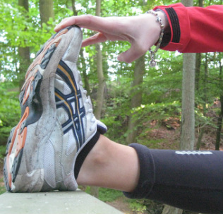
    One of the more common interventions that is performed is stretching. A study we looked at compared the results of the standard achilles tendon stretch to a sitting plantar fascia stretch. For the plantar fascia stretch, the patient would cross his/her legs and place the affected foot on the opposite knee. The patient then grasps the toes (especially the big toe) and maximally dorsiflexes them until a stretch is felt in the foot. In the study, the patients would perform their stretch first thing in the morning and before getting up after sitting for awhile. The study found both interventions to be successful, but the plantar fascia specific stretch more so. It should be noted that the study had no true control to rule out the patients' improvements due to natural healing processes.
   
    Dry needling is still a limited treatment technique for physical therapists; however, patients can have access to acupuncture on a wider basis. One article compared two groups to see the effect of acupuncture on plantar fasciitis. Both groups received standard treatments, such as icing, stretching, intrinsic foot strengthening, and NSAIDs. One group received acupuncture, additionally. Both groups found improvements in pain. There was no difference between the two groups after 4 weeks, but the acupuncture group was slightly better after 8 weeks.
   
Picture
    A treatment technique that is gaining popularity involves Instrument Assisted Soft Tissue Mobilization (IASTM). There are many products out there that fall under the category of IASTM: Graston Technique, ASTYM, Edge Tool, etc. The theory is generally the same behind them in that, through use of the tools, a healing inflammatory phase can be initiated by stimulating blood flow, nutrients, and fibroblasts to the area. Through proliferation of the fibroblasts, healing and formation of collagen can begin. The soft tissue mobilization can additionally aide in reorganization of the collagen fibers to proper orientation. This study in particular was a preliminary look at Graston Technique, discussing the theory, protocol, initial evidence, and some case studies. As plantar fasciitis is a soft tissue pathology, IASTM could have useful implications for patients with this disorder. When further research is performed on the subject, it may be found that IASTM has a very important place in treating these patients.
References:
Abd El Salam MS, Abd Elhafz YN. "Low-dye taping versus medial arch support in managing pain and pain-related disability in patients with plantar fasciitis." Foot Ankle Spec. 2011 Apr;4(2):86-91. Web. 10/13/12.

Bassford, Jeffrey. "A Randomized Controlled Evaluation of Low Level Laser Therapy: Plantar Fasciitis." Arch Phys Med Rehabil. 79. (1998): n. page. Web. 8 Oct. 2012.

Beyzadeoğlu T, Gökçe A, Bekler H. "[The effectiveness of dorsiflexion night splint added to conservative treatment for plantar fasciitis]." Acta Orthop Traumatol Turc. 2007;41(3):220-4. Web. 10/13/12.
Digiovanni BF
, Nawoczenski DA, Malay DP, Graci PA, Williams TT, Wilding GE, Baumhauer JF. "Plantar fascia-specific stretching exercise improves outcomes in patients with chronic plantar fasciitis. A prospective clinical trial with two-year follow-up." J Bone Joint Surg Am. 2006 Aug;88(8):1775-81. Web. 10/12/12.

Hammer, WI. "The effect of mechanical load on degenerated soft tissue." J body Mov Ther. 12.3 (2008): 246-256. Web. 8 Oct. 2012.

Hyland, Matthew. "Randomized Control Trial of Calcaneal Taping, Sham Taping, and Plantar Fascia Stretching for the Short-term Management of Plantar Heel Pain." Journal of Orthopaedic and Sports Physical Therapy. 36.6 (2006): n. page. Web. 8 Oct. 2012.

Karagounis P, Tsironi M, Prionas G, Tsiganos G, Baltopoulos P. "Treatment of plantar fasciitis in recreational athletes: two different therapeutic protocols." Foot Ankle Spec. 2011 Aug;4(4):226-34. Web. 10/13/12.

Lee SY, McKeon P, Hertel J. "Does the use of orthoses improve self-reported pain and function measures in patients with plantar fasciitis? A meta-analysis." Phys Ther Sport. 2009 Feb;10(1):12-8. Web. 10/10/2012.

Renan-Ording, Romulo. "Effectiveness of Myofascial Trigger Point Manual Therapy Combined with a Self-stretching Protocol for the Management of Plantar Heel Pain: A Randomized Control Trial." Journal of Orthopedic and Sports Physical Therapy. 41.2 (2011): 43-51. Web. 8 Oct. 2012.

Ryan, M, S Fraser, K McDonald, and J Taunton. "Examining the degree of pain reduction using a multielement exercise model with a conventional training shoe versus an ultraflexible training shoe for treating plantar fasciitis." Phys Sportsmed. 37.4 (2009): 67-84. Web. 10 Oct. 2012.

Stratton M, McPoil TG, Cornwall MW, Patrick K. "Use of low-frequency electrical stimulation for the treatment of plantar fasciitis." J Am Podiatr Med Assoc. 2009 Nov-Dec;99(6):481-8. Web. 10/13/12.
1 Comment
using essay writing services link
8/25/2022 07:09:09 am

Pretty useful update for the various people who are looking for the wise help here. I hope through the physical treatment on your forum the people able to understands the health problems. Do share more updates here must visit again to learn.

Reply


Leave a Reply.

    Picture
    Dr. Brian Schwabe's NEW Book in partner with PaleoHacks!
    Picture
    Learn residency-level content on our
    Insider Access pages
    Picture

    Picture

    We value quality PT education & CEU's.  Click the MedBridge logo below for TSPT savings!


    Archives

    July 2019
    June 2019
    May 2019
    March 2019
    February 2019
    January 2019
    December 2018
    November 2018
    October 2018
    September 2018
    August 2018
    July 2018
    June 2018
    May 2018
    April 2018
    March 2018
    February 2018
    January 2018
    December 2017
    November 2017
    October 2017
    September 2017
    August 2017
    July 2017
    June 2017
    May 2017
    April 2017
    March 2017
    February 2017
    January 2017
    December 2016
    November 2016
    October 2016
    September 2016
    August 2016
    July 2016
    June 2016
    May 2016
    April 2016
    March 2016
    February 2016
    January 2016
    December 2015
    November 2015
    October 2015
    September 2015
    August 2015
    July 2015
    June 2015
    May 2015
    April 2015
    March 2015
    February 2015
    January 2015
    December 2014
    November 2014
    October 2014
    September 2014
    August 2014
    July 2014
    June 2014
    May 2014
    April 2014
    March 2014
    February 2014
    January 2014
    December 2013
    November 2013
    October 2013
    September 2013
    August 2013
    July 2013
    June 2013
    May 2013
    April 2013
    March 2013
    February 2013
    January 2013
    December 2012
    November 2012
    October 2012
    September 2012
    August 2012

    Categories

    All
    Chest
    Core Muscle
    Elbow
    Foot
    Foot And Ankle
    Hip
    Knee
    Manual Therapy
    Modalities
    Motivation
    Neck
    Neural Tension
    Other
    Research
    Research Article
    Shoulder
    Sij
    Spine
    Sports
    Therapeutic Exercise


    RSS Feed

Home

Contact Us

Copyright © The Student Physical Therapist LLC 2022
Photos used under Creative Commons from dsearls, moo.review, liverpoolhls, matturick, lwpkommunikacio, Lake Clark NPS, Tobyotter, Charly Meunier, Vincent Albanese, CNBP, Zepfanman.com, aotaro, sportEX journals, jillccarlson, Yann07, US Department of Education, osseous, TheeErin, ajy591, Mark P / PIX41, Thomas Fisher Rare Book Library, UofT, teresatrimm, donnierayjones, NNelumba, Tobyotter, Frédéric de Villamil, Jordanhill School D&T Dept, danabooo, cadillacjr2002, ingridkreuz, RDECOM, U.S. Naval Forces Central Command/U.S. Fifth Fleet, Wonderlane, hectorir, Jeffrey, Chris Hunkeler, quillons, COD Newsroom, Alan Cleaver, CCFoodTravel.com, liverpoolhls, Dr.Farouk, Jeffrey, Sten Dueland, Håkan Dahlström, PPGWings, Parker Michael Knight, MLazarevski, dno1967b, Fimb, Jim Larrison, infomatique, U.S. Naval Forces Central Command/U.S. Fifth Fleet, Idhren, iwona_kellie, AnEternalGoldenBraid, quinn.anya, jlk.1, Villainette, mlsaero, roelandpype, Defence Images, CiscoANZ, istolethetv, Our Dream Photography (Personal), BraNewbs, Meg Stewart, phalinn, akeg, hectorir, philcampbell, MilitaryHealth, akeg, Army Medicine, Shar Ka, Subconsci Productions, Eric Kilby, Anonymous9000, quinn.anya, COD Newsroom, UMN Department of Plant Pathology, familymwr, all of olive., Keith Allison, JD Hancock, Synergy by Jasmine, roger_mommaerts, John-Morgan, a.drian, Instant Vantage, Beshef, markhillary, Vegar S Hansen Photography, DEC International, kaolin fire, smith_cl9, cinnamon_girl, jillccarlson, Extra Ketchup, brain_blogger, heyjoewhereyougoinwiththatguninyourhand, tv42, COMSALUD, Nicholas_T, Ano Lobb. @healthyrx, kaibara87, mlinksva, smbuckley23, sappsnap, Menage a Moi, brain_blogger, MaloMalverde, One Way Stock, ethet., ~ggvic~, MAClarke21, Keith Allison, pictures of money, mendolus shank, HotlantaVoyeur, Mr. T in DC, abbamouse, HystericalMark, Travis Hornung, jenny downing, shock264, Peter Mooney, Futurilla, sfslim, Emily Carlin, PFX Photo, ITU Pictures, TP studio, akunamatata, Magdalena Roeseler, osseous, smith_cl9, warrenski, Sh4rp_i, mrbichel, Renaud Camus, sicamp, GreenFlames09, infomatique, mark i geo, Volker Davids, TerryJohnston, Daniela Vladimirova, CJS*64, VarsityLife, Quentin Verwaerde, Annie Mole, US Army Africa, RLHyde, openBIT e. V., Vox Efx, fabola, Gerry Dincher, mlsaero, Infomastern, N4n0, Wonderlane, TheeErin, Nanagyei, gogogadgetscott, Pulmonary Pathology, PELeCON, Elvert Barnes, @RunRockPrincess, woodleywonderworks, Asbestos Bill, Joelk75, otisarchives4, jillccarlson, sportEX journals, The Wandering Angel, perpetualplum, V31S70, colecamp, jpalinsad360, JulyYu, Andres Rueda, Whenleavesfall, melloveschallah, Pink Sherbet Photography, canonim, BrianHenry ////|//, Leonard John Matthews, mikecogh, Zepfanman.com, Kevin M. Gill, Claire L. Evans, afunkydamsel, The Official CTBTO Photostream, cyOFdevelin fame, dok1, Pam loves pie, hoshi7, opensourceway
  • Home
  • About Us
  • Insider Access
    • About Insider Access
  • Online Courses
    • Hooper's Knee
    • Physical Therapist Entrepreneur Course
    • Physical Therapist Consultant Course
    • Orthopedic Management of the Cervical Spine
    • Return to Sport Essentials Course
  • Resources
    • Newsletter
    • Business Minded Sports Physio Podcast
    • Day in the Life of a Sports PT
    • Residency Corner
    • Special Tests >
      • Cervical Spine >
        • Alar Ligament Test
        • Bakody's Sign
        • Cervical Distraction Test
        • Cervical Rotation Lateral Flexion Test
        • Craniocervical Flexion Test (CCFT)
        • Deep Neck Flexor Endurance Test
        • Posterior-Anterior Segmental Mobility
        • Segmental Mobility
        • Sharp-Purser Test
        • Spurling's Maneuver
        • Transverse Ligament Test
        • ULNT - Median
        • ULNT - Radial
        • ULNT - Ulnar
        • Vertebral Artery Test
      • Thoracic Spine >
        • Adam's Forward Bend Test
        • Passive Neck Flexion Test
        • Thoracic Compression Test
        • Thoracic Distraction Test
        • Thoracic Foraminal Closure Test
      • Lumbar Spine/Sacroiliac Joint >
        • Active Sit-Up Test
        • Alternate Gillet Test
        • Crossed Straight Leg Raise Test
        • Extensor Endurance Test
        • FABER Test
        • Fortin's Sign
        • Gaenslen Test
        • Gillet Test
        • Gower's Sign
        • Lumbar Quadrant Test
        • POSH Test
        • Posteroanterior Mobility
        • Prone Knee Bend Test
        • Prone Instability Test
        • Resisted Abduction Test
        • Sacral Clearing Test
        • Seated Forward Flexion Test
        • SIJ Compression/Distraction Test
        • Slump Test
        • Sphinx Test
        • Spine Rotators & Multifidus Test
        • Squish Test
        • Standing Forward Flexion Test
        • Straight Leg Raise Test
        • Supine to Long Sit Test
      • Shoulder >
        • Active Compression Test
        • Anterior Apprehension
        • Biceps Load Test II
        • Drop Arm Sign
        • External Rotation Lag Sign
        • Hawkins-Kennedy Impingement Sign
        • Horizontal Adduction Test
        • Internal Rotation Lag Sign
        • Jobe Test
        • Ludington's Test
        • Neer Test
        • Painful Arc Sign
        • Pronated Load Test
        • Resisted Supination External Rotation Test
        • Speed's Test
        • Posterior Apprehension
        • Sulcus Sign
        • Thoracic Outlet Tests >
          • Adson's Test
          • Costoclavicular Brace
          • Hyperabduction Test
          • Roos (EAST)
        • Yergason's Test
      • Elbow >
        • Biceps Squeeze Test
        • Chair Sign
        • Cozen's Test
        • Elbow Extension Test
        • Medial Epicondylalgia Test
        • Mill's Test
        • Moving Valgus Stress Test
        • Push-up Sign
        • Ulnar Nerve Compression Test
        • Valgus Stress Test
        • Varus Stress Test
      • Wrist/Hand >
        • Allen's Test
        • Carpal Compression Test
        • Finkelstein Test
        • Phalen's Test
        • Reverse Phalen's Test
      • Hip >
        • Craig's Test
        • Dial Test
        • FABER Test
        • FAIR Test
        • Fitzgerald's Test
        • Hip Quadrant Test
        • Hop Test
        • Labral Anterior Impingement Test
        • Labral Posterior Impingement Test
        • Long-Axis Femoral Distraction Test
        • Noble Compression Test
        • Percussion Test
        • Sign of the Buttock
        • Trendelenburg Test
      • Knee >
        • Anterior Drawer Test
        • Dial Test (Tibial Rotation Test)
        • Joint Line Tenderness
        • Lachman Test
        • McMurray Test
        • Noble Compression Test
        • Pivot-Shift Test
        • Posterior Drawer Test
        • Posterior Sag Sign
        • Quad Active Test
        • Thessaly Test
        • Valgus Stress Test
        • Varus Stress Test
      • Foot/Ankle >
        • Anterior Drawer
        • Calf Squeeze Test
        • External Rotation Test
        • Fracture Screening Tests
        • Impingement Sign
        • Navicular Drop Test
        • Squeeze Test
        • Talar Tilt
        • Tarsal Tunnel Syndrome Test
        • Test for Interdigital Neuroma
        • Windlass Test