The Student​ Physical Therapist
  • Home
    • Newsletter
    • About
  • Insider Access
    • About Insider Access
  • Brian's Corner
    • Sports & Entrepreneurship Blog
    • Return to Sport Tests
    • PT Entrepreneur Course
    • Return to Sport Essentials Course
    • Become a PAID PT Consultant Course
  • Chris' Corner
  • Jim's Corner
    • Orthopedic Blog
  • Special Tests
    • Cervical Spine >
      • Alar Ligament Test
      • Bakody's Sign
      • Cervical Distraction Test
      • Cervical Rotation Lateral Flexion Test
      • Craniocervical Flexion Test (CCFT)
      • Deep Neck Flexor Endurance Test
      • Posterior-Anterior Segmental Mobility
      • Segmental Mobility
      • Sharp-Purser Test
      • Spurling's Maneuver
      • Transverse Ligament Test
      • ULNT - Median
      • ULNT - Radial
      • ULNT - Ulnar
      • Vertebral Artery Test
    • Thoracic Spine >
      • Adam's Forward Bend Test
      • Passive Neck Flexion Test
      • Thoracic Compression Test
      • Thoracic Distraction Test
      • Thoracic Foraminal Closure Test
    • Lumbar Spine/Sacroiliac Joint >
      • Active Sit-Up Test
      • Alternate Gillet Test
      • Crossed Straight Leg Raise Test
      • Extensor Endurance Test
      • FABER Test
      • Fortin's Sign
      • Gaenslen Test
      • Gillet Test
      • Gower's Sign
      • Lumbar Quadrant Test
      • POSH Test
      • Posteroanterior Mobility
      • Prone Knee Bend Test
      • Prone Instability Test
      • Resisted Abduction Test
      • Sacral Clearing Test
      • Seated Forward Flexion Test
      • SIJ Compression/Distraction Test
      • Slump Test
      • Sphinx Test
      • Spine Rotators & Multifidus Test
      • Squish Test
      • Standing Forward Flexion Test
      • Straight Leg Raise Test
      • Supine to Long Sit Test
    • Shoulder >
      • Active Compression Test
      • Anterior Apprehension
      • Biceps Load Test II
      • Drop Arm Sign
      • External Rotation Lag Sign
      • Hawkins-Kennedy Impingement Sign
      • Horizontal Adduction Test
      • Internal Rotation Lag Sign
      • Jobe Test
      • Ludington's Test
      • Neer Test
      • Painful Arc Sign
      • Pronated Load Test
      • Resisted Supination External Rotation Test
      • Speed's Test
      • Posterior Apprehension
      • Sulcus Sign
      • Thoracic Outlet Tests >
        • Adson's Test
        • Costoclavicular Brace
        • Hyperabduction Test
        • Roos (EAST)
      • Yergason's Test
    • Elbow >
      • Biceps Squeeze Test
      • Chair Sign
      • Cozen's Test
      • Elbow Extension Test
      • Medial Epicondylalgia Test
      • Mill's Test
      • Moving Valgus Stress Test
      • Push-up Sign
      • Ulnar Nerve Compression Test
      • Valgus Stress Test
      • Varus Stress Test
    • Wrist/Hand >
      • Allen's Test
      • Carpal Compression Test
      • Finkelstein Test
      • Phalen's Test
      • Reverse Phalen's Test
    • Hip >
      • Craig's Test
      • Dial Test
      • FABER Test
      • FAIR Test
      • Fitzgerald's Test
      • Hip Quadrant Test
      • Hop Test
      • Labral Anterior Impingement Test
      • Labral Posterior Impingement Test
      • Long-Axis Femoral Distraction Test
      • Noble Compression Test
      • Percussion Test
      • Sign of the Buttock
      • Trendelenburg Test
    • Knee >
      • Anterior Drawer Test
      • Dial Test (Tibial Rotation Test)
      • Joint Line Tenderness
      • Lachman Test
      • McMurray Test
      • Noble Compression Test
      • Pivot-Shift Test
      • Posterior Drawer Test
      • Posterior Sag Sign
      • Quad Active Test
      • Thessaly Test
      • Valgus Stress Test
      • Varus Stress Test
    • Foot/Ankle >
      • Anterior Drawer
      • Calf Squeeze Test
      • External Rotation Test
      • Fracture Screening Tests
      • Impingement Sign
      • Navicular Drop Test
      • Squeeze Test
      • Talar Tilt
      • Tarsal Tunnel Syndrome Test
      • Test for Interdigital Neuroma
      • Windlass Test
    • HEP >
      • Neck and Shoulder >
        • Supine Chin Tuck
        • Supine DNF with Towel Assist
        • Supine DNF
        • Standing Chin Tuck Against Wall
        • Standing Chin Tuck Against Wall with Scaption
        • Seated Cervical Retraction Repeated
        • Seated Cervical Retraction with Extension Repeated
        • Seated Cervical Retraction with Sidebend Repeated
        • Seated Cervical Retraction with Rotation Repeated
        • Standing Wall Shrugs at 90 Degrees Flex
        • Seated Thoracic Whips
        • Standing Ballistic Shoulder Extensions
        • Standing Repeated Shoulder Extension with Squat
        • Standing Repetead Shoulder Horiz. Abd. with Ext. CKC
        • Seated with Arms on Pillows Cervical AROM (Flex/Ext/Rot/SB)
        • Seated with Arms on Pillows Shrugs
        • Seated with Arms on Pillows Shrug with Scapular Retraction
        • Supine Shoulder IR with GH Centralization
        • Supine Shoulder ER with GH Centralization
        • Holding Dumbbell at 180 Degrees Flexion for Time
        • Cat Camel
        • Prone T's
        • Prone Y's
        • Quad Chin Tuck w/ Shoulder Flexion
      • Low Back >
        • Supine TA Isometric
        • Standing TA Isometric Agains Wall with Squat
        • Supine BKFO
        • Quad Rock Back
        • Standing Hip Hinge
        • Sit to Stand with Hip Hinge
        • Repeated Lumbar Sideglides
        • Repeated Standing Lumbar Extension
        • Repeated Standing Lumbar Flexion
        • Repeated Prone Press-Ups
        • Repeated Supine DKC
        • Slump Sciatic Nerve Glides
        • Birddog Progression
      • Hip and Knee >
        • Clamshells with Progressions
        • Fire Hydrants with Progressions
        • Donkey Kicks
        • Bridge Variations
        • Repeated Hip Flexion
        • Squats
        • Seated Repeated Knee Extensions
        • CKC Seated Repeated Knee Extensions
        • Heel Slides
        • CKC DF with Tibial IR
      • Foot and Ankle >
        • Calf Raises
        • Calf Raises with Soccer Ball Between Medial Malleoli
        • Towel Scrunches with Foot in PF
        • Toe Flexion Using T-Band with Foot in PF
        • PF with Toes Flexed Using T-Band
        • DF with Toes Flexed Using T-Band
        • Forefoot Adduction
        • Gastroc Stretch
        • Repeated PF
      • Examination Templates
Picture

Plica of the Knee: What is it and how do you treat it?

5/22/2013

0 Comments

 
Picture
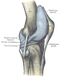
Plica! I am sure you have heard of them, but do you know the likelihood of one of these patients showing up at your clinic door? Should you consider any special treatment recommendations with this population? Keep reading to find out!

Plica are inward folds of the synovial lining of the knee. Because they are embryonic deviations, plica are seen inconsistently throughout the population. Typically, they do not cause any pain and are asymptomatic, but they can become inflamed resulting in a "plica syndrome." From a clinical standpoint, it is important to note that "symptoms...are indistinguishable from other intra-articular conditions such as meniscal tears, articular cartilage injuries, or osteochondritic lesions, creating a diagnostic conundrum." 

There are 4 general categories of plica: suprapatellar, medial parapatellar, lateral, and infrapatellar. Lateral plica are very rarely seen and consequently little research has been conducted on them. Classification of each other type of plica becomes confusing because each one has multiple sub-groups. It is important to know that the suprapatellar plica does have attachments to the quadriceps tendon and depending on the size and shape, may create impingement between the patellar tendon and femoral trochlea in the range of 70-100 degrees of knee flexion. Similarly, the medial patellar plica also changes orientation with different positions of the knee. Its origin and attachment is controversial with several authors listing different anatomic variations. Finally, the infrapatellar plica is the most common plica in the population with 85% of patients presenting with one according to a study by Wachter in 1979. It runs superior-inferior from the intercondylar notch into the infrapatellar fat pad. The incidence of individuals with plica syndrome is under controversy, and some surgeons believe it is over-diagnosed. 

Clinically, 50% of patient's with plica syndrome will have a history of trauma or twisting at the knee joint. Once the initial injury is resolved, a patient maybe asymptomatic for a period of time, only to report back to the clinic weeks or months later with intense anterior knee pain. Anterior knee pain is known as the cardinal symtpom regardless of which plica is affected. Additionally, they may have complaints of a clicking or popping in the knee with flexion and describe the pain as intermittent, dull, and achy. Others report a tightness around the anterior medial knee joint which increases with deeper knee flexion angles. Because the incidence of plica syndrome is low, diligent differential diagnosis is a must, ruling out other possible causes of anterior knee pain. A clinical diagnosis alone is very difficult. Recent studies have shown the both MRI and Ultrasound have had good success in viewing plica shape and size.

So what can we do for these patients? Unfortunately, "success rates of conservative management are generally low" with age being a predictive factor for your success. The younger patient is more likely to respond favorably to conservative treatment since they have not suffered the long-term effects of impinging plica and resulting structural changes within the knee joint. Therapy should consist of a period of rest from deep knee flexion activites, followed by a course of NSAIDs to help curb inflammation. Caution should be taken with intense exercise progression secondary to likelihood of aggravating the plica. Additionally, success rates of PT alone are variable and surgical intervention may be required with persistent symptoms. It should be noted that while this is a recent article, many of the references under conservative management are fairly dated. The therapy profession has since grown in its knowledge of conservative treatment for knee conditions and the importance of regional interdependence around the knee joint. 

In conclusion, plica syndome is not something you will see in the clinic everyday, but having this knowledge in your toolbox will make you a more well-rounded clinician. Reading this article would be very beneficial because there are great pictures discussing different variations and anatomic location.

References: 
Schindler O. 2013. 'The Sneaky Plica' revisited: morphology, pathophysiology, and treatment of synovial plicae of the knee. Knee Surg Sports Traumatol Arthrosc. 2013; 2013. Web. 14 May 2013. 
0 Comments


Leave a Reply.

    Picture
    Dr. Brian Schwabe's NEW Book in partner with PaleoHacks!
    Picture
    Learn residency-level content on our
    Insider Access pages
    Picture

    Picture

    We value quality PT education & CEU's.  Click the MedBridge logo below for TSPT savings!


    Archives

    July 2019
    June 2019
    May 2019
    March 2019
    February 2019
    January 2019
    December 2018
    November 2018
    October 2018
    September 2018
    August 2018
    July 2018
    June 2018
    May 2018
    April 2018
    March 2018
    February 2018
    January 2018
    December 2017
    November 2017
    October 2017
    September 2017
    August 2017
    July 2017
    June 2017
    May 2017
    April 2017
    March 2017
    February 2017
    January 2017
    December 2016
    November 2016
    October 2016
    September 2016
    August 2016
    July 2016
    June 2016
    May 2016
    April 2016
    March 2016
    February 2016
    January 2016
    December 2015
    November 2015
    October 2015
    September 2015
    August 2015
    July 2015
    June 2015
    May 2015
    April 2015
    March 2015
    February 2015
    January 2015
    December 2014
    November 2014
    October 2014
    September 2014
    August 2014
    July 2014
    June 2014
    May 2014
    April 2014
    March 2014
    February 2014
    January 2014
    December 2013
    November 2013
    October 2013
    September 2013
    August 2013
    July 2013
    June 2013
    May 2013
    April 2013
    March 2013
    February 2013
    January 2013
    December 2012
    November 2012
    October 2012
    September 2012
    August 2012

    Categories

    All
    Chest
    Core Muscle
    Elbow
    Foot
    Foot And Ankle
    Hip
    Knee
    Manual Therapy
    Modalities
    Motivation
    Neck
    Neural Tension
    Other
    Research
    Research Article
    Shoulder
    Sij
    Spine
    Sports
    Therapeutic Exercise


    RSS Feed

Home

Contact Us

Copyright © The Student Physical Therapist LLC 2020
Photos used under Creative Commons from dsearls, moo.review, liverpoolhls, matturick, lwpkommunikacio, Lake Clark NPS, Tobyotter, Charly Meunier, Vincent Albanese, CNBP, Zepfanman.com, aotaro, sportEX journals, jillccarlson, Yann07, US Department of Education, osseous, TheeErin, ajy591, Mark P / PIX41, Thomas Fisher Rare Book Library, UofT, teresatrimm, donnierayjones, NNelumba, Tobyotter, Frédéric de Villamil, Jordanhill School D&T Dept, danabooo, cadillacjr2002, ingridkreuz, RDECOM, U.S. Naval Forces Central Command/U.S. Fifth Fleet, Wonderlane, hectorir, Jeffrey, Chris Hunkeler, quillons, COD Newsroom, Alan Cleaver, CCFoodTravel.com, liverpoolhls, Dr.Farouk, Jeffrey, Sten Dueland, Håkan Dahlström, PPGWings, Parker Michael Knight, MLazarevski, dno1967b, Fimb, Jim Larrison, infomatique, U.S. Naval Forces Central Command/U.S. Fifth Fleet, Idhren, iwona_kellie, AnEternalGoldenBraid, quinn.anya, jlk.1, Villainette, mlsaero, roelandpype, Defence Images, CiscoANZ, istolethetv, Our Dream Photography (Personal), BraNewbs, Meg Stewart, phalinn, akeg, hectorir, philcampbell, MilitaryHealth, akeg, Army Medicine, Shar Ka, Subconsci Productions, Eric Kilby, Anonymous9000, quinn.anya, COD Newsroom, UMN Department of Plant Pathology, familymwr, all of olive., Keith Allison, JD Hancock, Synergy by Jasmine, roger_mommaerts, John-Morgan, a.drian, Instant Vantage, Beshef, markhillary, Vegar S Hansen Photography, DEC International, kaolin fire, smith_cl9, cinnamon_girl, jillccarlson, Extra Ketchup, brain_blogger, heyjoewhereyougoinwiththatguninyourhand, tv42, COMSALUD, Nicholas_T, Ano Lobb. @healthyrx, kaibara87, mlinksva, smbuckley23, sappsnap, Menage a Moi, brain_blogger, MaloMalverde, One Way Stock, ethet., ~ggvic~, MAClarke21, Keith Allison, pictures of money, mendolus shank, HotlantaVoyeur, Mr. T in DC, abbamouse, HystericalMark, Travis Hornung, jenny downing, shock264, Peter Mooney, Futurilla, sfslim, Emily Carlin, PFX Photo, ITU Pictures, TP studio, akunamatata, Magdalena Roeseler, osseous, smith_cl9, warrenski, Sh4rp_i, mrbichel, Renaud Camus, sicamp, GreenFlames09, infomatique, mark i geo, Volker Davids, TerryJohnston, Daniela Vladimirova, CJS*64, VarsityLife, Quentin Verwaerde, Annie Mole, US Army Africa, RLHyde, openBIT e. V., Vox Efx, fabola, Gerry Dincher, mlsaero, Infomastern, N4n0, Wonderlane, TheeErin, Nanagyei, gogogadgetscott, Pulmonary Pathology, PELeCON, Elvert Barnes, @RunRockPrincess, woodleywonderworks, Asbestos Bill, Joelk75, otisarchives4, jillccarlson, sportEX journals, The Wandering Angel, perpetualplum, V31S70, colecamp, jpalinsad360, JulyYu, Andres Rueda, Whenleavesfall, melloveschallah, Pink Sherbet Photography, canonim, BrianHenry ////|//, Leonard John Matthews, mikecogh, Zepfanman.com, Kevin M. Gill, Claire L. Evans, afunkydamsel, The Official CTBTO Photostream, cyOFdevelin fame, dok1, Pam loves pie, hoshi7, opensourceway
  • Home
    • Newsletter
    • About
  • Insider Access
    • About Insider Access
  • Brian's Corner
    • Sports & Entrepreneurship Blog
    • Return to Sport Tests
    • PT Entrepreneur Course
    • Return to Sport Essentials Course
    • Become a PAID PT Consultant Course
  • Chris' Corner
  • Jim's Corner
    • Orthopedic Blog
  • Special Tests
    • Cervical Spine >
      • Alar Ligament Test
      • Bakody's Sign
      • Cervical Distraction Test
      • Cervical Rotation Lateral Flexion Test
      • Craniocervical Flexion Test (CCFT)
      • Deep Neck Flexor Endurance Test
      • Posterior-Anterior Segmental Mobility
      • Segmental Mobility
      • Sharp-Purser Test
      • Spurling's Maneuver
      • Transverse Ligament Test
      • ULNT - Median
      • ULNT - Radial
      • ULNT - Ulnar
      • Vertebral Artery Test
    • Thoracic Spine >
      • Adam's Forward Bend Test
      • Passive Neck Flexion Test
      • Thoracic Compression Test
      • Thoracic Distraction Test
      • Thoracic Foraminal Closure Test
    • Lumbar Spine/Sacroiliac Joint >
      • Active Sit-Up Test
      • Alternate Gillet Test
      • Crossed Straight Leg Raise Test
      • Extensor Endurance Test
      • FABER Test
      • Fortin's Sign
      • Gaenslen Test
      • Gillet Test
      • Gower's Sign
      • Lumbar Quadrant Test
      • POSH Test
      • Posteroanterior Mobility
      • Prone Knee Bend Test
      • Prone Instability Test
      • Resisted Abduction Test
      • Sacral Clearing Test
      • Seated Forward Flexion Test
      • SIJ Compression/Distraction Test
      • Slump Test
      • Sphinx Test
      • Spine Rotators & Multifidus Test
      • Squish Test
      • Standing Forward Flexion Test
      • Straight Leg Raise Test
      • Supine to Long Sit Test
    • Shoulder >
      • Active Compression Test
      • Anterior Apprehension
      • Biceps Load Test II
      • Drop Arm Sign
      • External Rotation Lag Sign
      • Hawkins-Kennedy Impingement Sign
      • Horizontal Adduction Test
      • Internal Rotation Lag Sign
      • Jobe Test
      • Ludington's Test
      • Neer Test
      • Painful Arc Sign
      • Pronated Load Test
      • Resisted Supination External Rotation Test
      • Speed's Test
      • Posterior Apprehension
      • Sulcus Sign
      • Thoracic Outlet Tests >
        • Adson's Test
        • Costoclavicular Brace
        • Hyperabduction Test
        • Roos (EAST)
      • Yergason's Test
    • Elbow >
      • Biceps Squeeze Test
      • Chair Sign
      • Cozen's Test
      • Elbow Extension Test
      • Medial Epicondylalgia Test
      • Mill's Test
      • Moving Valgus Stress Test
      • Push-up Sign
      • Ulnar Nerve Compression Test
      • Valgus Stress Test
      • Varus Stress Test
    • Wrist/Hand >
      • Allen's Test
      • Carpal Compression Test
      • Finkelstein Test
      • Phalen's Test
      • Reverse Phalen's Test
    • Hip >
      • Craig's Test
      • Dial Test
      • FABER Test
      • FAIR Test
      • Fitzgerald's Test
      • Hip Quadrant Test
      • Hop Test
      • Labral Anterior Impingement Test
      • Labral Posterior Impingement Test
      • Long-Axis Femoral Distraction Test
      • Noble Compression Test
      • Percussion Test
      • Sign of the Buttock
      • Trendelenburg Test
    • Knee >
      • Anterior Drawer Test
      • Dial Test (Tibial Rotation Test)
      • Joint Line Tenderness
      • Lachman Test
      • McMurray Test
      • Noble Compression Test
      • Pivot-Shift Test
      • Posterior Drawer Test
      • Posterior Sag Sign
      • Quad Active Test
      • Thessaly Test
      • Valgus Stress Test
      • Varus Stress Test
    • Foot/Ankle >
      • Anterior Drawer
      • Calf Squeeze Test
      • External Rotation Test
      • Fracture Screening Tests
      • Impingement Sign
      • Navicular Drop Test
      • Squeeze Test
      • Talar Tilt
      • Tarsal Tunnel Syndrome Test
      • Test for Interdigital Neuroma
      • Windlass Test
    • HEP >
      • Neck and Shoulder >
        • Supine Chin Tuck
        • Supine DNF with Towel Assist
        • Supine DNF
        • Standing Chin Tuck Against Wall
        • Standing Chin Tuck Against Wall with Scaption
        • Seated Cervical Retraction Repeated
        • Seated Cervical Retraction with Extension Repeated
        • Seated Cervical Retraction with Sidebend Repeated
        • Seated Cervical Retraction with Rotation Repeated
        • Standing Wall Shrugs at 90 Degrees Flex
        • Seated Thoracic Whips
        • Standing Ballistic Shoulder Extensions
        • Standing Repeated Shoulder Extension with Squat
        • Standing Repetead Shoulder Horiz. Abd. with Ext. CKC
        • Seated with Arms on Pillows Cervical AROM (Flex/Ext/Rot/SB)
        • Seated with Arms on Pillows Shrugs
        • Seated with Arms on Pillows Shrug with Scapular Retraction
        • Supine Shoulder IR with GH Centralization
        • Supine Shoulder ER with GH Centralization
        • Holding Dumbbell at 180 Degrees Flexion for Time
        • Cat Camel
        • Prone T's
        • Prone Y's
        • Quad Chin Tuck w/ Shoulder Flexion
      • Low Back >
        • Supine TA Isometric
        • Standing TA Isometric Agains Wall with Squat
        • Supine BKFO
        • Quad Rock Back
        • Standing Hip Hinge
        • Sit to Stand with Hip Hinge
        • Repeated Lumbar Sideglides
        • Repeated Standing Lumbar Extension
        • Repeated Standing Lumbar Flexion
        • Repeated Prone Press-Ups
        • Repeated Supine DKC
        • Slump Sciatic Nerve Glides
        • Birddog Progression
      • Hip and Knee >
        • Clamshells with Progressions
        • Fire Hydrants with Progressions
        • Donkey Kicks
        • Bridge Variations
        • Repeated Hip Flexion
        • Squats
        • Seated Repeated Knee Extensions
        • CKC Seated Repeated Knee Extensions
        • Heel Slides
        • CKC DF with Tibial IR
      • Foot and Ankle >
        • Calf Raises
        • Calf Raises with Soccer Ball Between Medial Malleoli
        • Towel Scrunches with Foot in PF
        • Toe Flexion Using T-Band with Foot in PF
        • PF with Toes Flexed Using T-Band
        • DF with Toes Flexed Using T-Band
        • Forefoot Adduction
        • Gastroc Stretch
        • Repeated PF
      • Examination Templates